Answering your questions: Adult-onset neurodegenerative diseases in dogs and cats
Neurodegenerative diseases in dogs and cats can result from a wide variety of pathologic processes.
Q: How can we better identify adult-onset neurodegenerative diseases in our patients?
Although many neurodegenerative diseases occur in dogs and cats, most of the well-described ones occur in neonatal or juvenile animals (e.g. cerebellar abiotrophies, most lysosomal storage diseases).1 In diagnosing adult-onset neurodegenerative disorders, two problems are seen. First, there may be a tendency to overdiagnose disorders such as degenerative myelopathy and canine cognitive dysfunction. This may occur because many owners of aged animals decline a full workup to avoid the anesthetic risk and are reluctant to intervene therapeutically in older animals. Because they choose not to intervene, owners more readily accept a diagnosis of a disease for which little can be done.
Second, practitioners may be uncomfortable diagnosing neurodegenerative disease in adult animals because relatively few adult-onset neurodegenerative disorders are well-described and, therefore, recognized by veterinarians. In the past, subtle changes in central nervous system (CNS) morphology were not identified on computed tomography images. The advent of magnetic resonance imaging (MRI) has changed this. During the next decade, new disease entities will be described.
When a neurodegenerative disease is suspected
1. Identify and document neurologic deficits.
2. Conduct a full diagnostic workup in all animals with progressive neurologic signs.
3. Stay informed of advances in veterinary neurology, particularly as MRI becomes more accessible in dogs and cats.
Q: Which neurodegenerative diseases have an adult onset in dogs and cats?
Neurodegenerative diseases in dogs and cats can result from a wide variety of pathologic processes. These can be grouped as inherited disorders, in which an inborn defect causes progressive neuronal loss, and as acquired disorders that reflect an accumulation of environmental insults over time (Table 1).

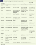
Table 1. Adult-onset Neurodegenerative Diseases in Small Animals
Inherited disorders
The best-described inherited neurodegenerative diseases are the lysosomal storage diseases. Although most of these affect neonatal and juvenile animals, some storage diseases have a late onset. These include mucopolysaccharidosis type IIIB in Schipperkes and ceroid lipofuscinosis. The former disease causes progressive cerebellar signs in Schipperkes, first evident between 3 and 5 years of age.2 Because of the insidious nature of the signs, the animal may not be presented to a veterinarian until months after disease onset. Ceroid lipofuscinosis is a disease process distinct from the intraneuronal accumulation of lipofuscin that occurs with age.3 This storage disorder has been recognized in Tibetan terriers, English setters, cocker spaniels, dachshunds, and border collies, with sporadic reports in many other dog and cat breeds. Clinical signs usually develop in animals younger than 2 years old, but an onset of as late as 9 years of age has been reported, particularly in dachshunds.3 Signs usually reflect forebrain and retinal involvement, with changes in behavior and loss of night vision (nyctalopia) predominating. However, the changes have a diffuse distribution, and a wide range of signs have been reported including ataxia, paresis, head tremors, and seizures.
Inherited cerebellar degenerative disorders with an adult onset have been described in American Staffordshire terriers4 and related American pit bull terriers as well as Brittany spaniels.5 In these breeds, an onset of cerebellar and vestibular signs (e.g. hypermetria, ataxia, nystagmus) occurs around 4 to 6 years of age. The signs are slowly progressive, and as a result, affected animals may be presented to their veterinarians late in life. Other breeds with inherited cerebellar degeneration that can present in adulthood include Gordon setters,6 Old English sheepdogs,7 and Scottish terriers.8 In these breeds, however, signs usually first appear when the animal is still a juvenile (6 to 18 months).
Breed-related disorders suspected to be inherited
The next group of diseases includes those thought to be inherited because of an established breed predisposition but for which a mode of inheritance has not been determined. The best known of these is degenerative myelopathy. This disease is well-described in German shepherds and occurs sporadically in other large breeds.9 It appears to be an emerging problem in Pembroke Welsh corgis, boxers, and Rhodesian ridgebacks, although these breeds may be suffering from a different degenerative disease process than that seen in German shepherds. Degenerative myelopathy causes progressive paraparesis in dogs older than 5 years of age. Usually the affected dogs are 9 years or older.
There are isolated reports of rare breed-related neurodegenerative diseases in dogs with an onset of signs well into adulthood. Rottweilers suffer from several breed-related neurodegenerative diseases, but the only one with a true adult onset of signs is leukoencephalomyelopathy.10 Progressive signs of ataxia, hypermetria, and tetraparesis appear between 1 and 4 years of age and are slowly progressive. In addition, neuroaxonal dystrophy in rottweilers11 usually results in signs of tetraparesis before 1 year of age; however, because the signs progress slowly over a number of years, this disease should also be considered in adult rottweilers. A necrotizing encephalopathy that has been likened to the human mitochondrial disorder known as Leigh's disease has been reported in five Alaskan huskies.12 In four of the affected dogs, an acute onset of ataxia, seizures, behavioral changes, and signs of cranial nerve deficits occurred before 1 year of age; however, in one dog the signs did not appear until 2.5 years of age.
Peripheral neuropathies characterized by degenerative changes may occur in older dogs.13 These include dancing Doberman disease, a degenerative neuropathy that affects the pelvic limbs of adult Doberman pinschers causing flexion of the limbs and a repeated shifting of weight, which give the appearance of dancing. This disease has a reported onset of signs as late as 7 years. Adult-onset polyneuropathies in Leonbergers, rottweilers, and Great Danes also cause generalized weakness.
Acquired diseases
The degenerative neuropathy associated with diabetes is well-recognized in cats and probably occurs subclinically in dogs. Insulinomas also cause a degenerative neuropathy in dogs, and hypothyroidism can cause generalized weakness due to a neuropathy.
Dysautonomia refers to a degenerative disease of the autonomic nervous system in dogs and cats that usually affects adult animals.14 Progressive loss of autonomic neurons causes a wide variety of debilitating autonomic signs, including mydriasis, keratoconjunctivitis sicca, regurgitation, urinary retention, constipation, and diarrhea; it is often fatal. Many theories exist about the cause, but increasing evidence suggests that, at least in cats, this disease is the result of Clostridium botulinum C toxin.15
Other disorders
Cognitive dysfunction
The less well characterized diseases are those that seem to be related to aging itself. All companion-animal veterinarians are familiar with an older dog or cat that, according to the owner, appears to have become senile. Signs can include a loss of hearing and vision, getting lost in familiar environments, a loss of housetraining, constant pacing, and a change in sleep patterns. In dogs, this aging syndrome has been called canine cognitive dysfunction, although many neurologists and behaviorists are still uncomfortable with the actual definition of the disease and what it encompasses.16
In experimental colonies of beagles, careful studies of learning ability and memory have shown that cognitive function declines with age.17 As would be expected, some dogs experience a more severe decline than others, and the most severely affected dogs have been compared to people with Alzheimer's disease. As cognitive testing has not been standardized in pet dogs, the diagnosis of canine cognitive dysfunction in this group depends on animals showing one or more of the following changes: disorientation, disruption of activity and sleep, changes in housetraining, and changes in interactions with family members.16 The older the dog, the more likely it is to have abnormalities in one or more category. However, because older dogs are more likely to also have orthopedic, metabolic, and cardiovascular disease, all of which could affect behavior in these categories, it is difficult to determine the extent to which neurodegenerative disease is contributing to the clinical signs.
Far less information is published about cognitive dysfunction in cats. Experimental behavioral studies in aged cats also suggest that their cognitive function declines.18
Cerebral microvascular disease
Cerebral microvascular disease is the second most common cause of cerebral neurodegeneration in people, producing a disease called vascular dementia.19 Although dogs and cats do not suffer from vascular disease to the same extent as people do, the advent of MRI has improved our ability to diagnose CNS vascular diseases. This area is still emerging in veterinary medicine. No published reports exist of vascular dementia in dogs and cats that corresponds to the disease in people. Focal large infarcts have been described in dogs and cats for many years (see boxed text titled "Do strokes occur in dogs and cats?").20 In both species, large strokes affecting the forebrain can result in persistent behavioral changes, but this is a distinct process when compared with the insidious loss of neurons secondary to repeated microvascular events as seen in people. As more aged dogs and cats undergo MRI studies of their brains, descriptions of changes that represent microvascular disease may emerge.


Do strokes occur in dogs and cats?
Q: What types of age-related changes occur in the canine brain?
The histopathologic changes associated with aging have been described in a number of studies in dogs. Beta-amyloid protein accumulates in diffuse deposits in the prefrontal cortex, entorhinal cortex, and hippocampus. The deposits are detected first in the prefrontal cortex, and their principal constituent is the β-amyloid 1-42 protein, the insoluble fragment that is thought to be neurotoxic.21 Apoptosis of neurons is also detected and tends to localize to those areas where amyloid accumulation is most prevalent.22 Evidence for progressive accumulation of oxidative damage also exists.23 These changes have been likened to the early changes seen in the brains of patients with Alzheimer's disease. Unlike Alzheimer's disease patients, however, dogs do not develop mature amyloid plaques with cores and do not develop neurofibrillary tangles.
The cerebral vasculature also shows pathologic changes with age. A shorter fragment of β-amyloid protein, the β-amyloid 1-40 fragment, accumulates in the walls of blood vessels, although the significance of these changes is not known.24 Dogs with hypothyroidism may develop cerebrovascular atherosclerosis, but unlike in people, this is not a change seen simply with age in dogs.25
Other reported aging changes include loss of spiral ganglion neurons in the inner ear associated with deafness26 and diffuse gliosis.27 Myelinated tracts develop nonspecific changes with age; in dogs over 12 years of age, white matter tracts, such as the corona radiata and corpus callosum, develop pallor and gliosis with accumulation of perivascular macrophages.28
Intracytoplasmic neuronal inclusions called Lafora's bodies are encountered in the brains of aged dogs, their frequency apparently correlating to age. Finally, accumulation of a pigment called lipofuscin is a common event in numerous organs, including those in the CNS. The pigment is brown and granular and tends to lie eccentrically between the nucleus and axon hillock of neuronal cell bodies. In severely affected neurons, it may be perinuclear or fill the entire cytoplasm. Less dense deposits may be found in astrocytes and oligodendrocytes.29
Changes in the aging canine brain have also been described on magnetic resonance images.30,31 There is progressive cortical atrophy and ventricular enlargement as dogs age. The brain blood volume does not change with age, and a nonsignificant trend toward increasing blood-brain barrier permeability exists.30 A detailed study revealed that the frontal lobe of the canine brain atrophies earlier than the rest of the brain.31
Q: What is the age range for the onset of these changes in dogs? How do they manifest clinically?
Histopathologic changes are seen as early as 2 to 3 years of age (e.g. lipofuscin accumulation) and accumulate throughout life.29 Frontal lobe atrophy was detected in beagles aged 8 years and older, perhaps correlating with the age at which β-amyloid deposits appear. Ventricular volume (as an indicator of cerebral atrophy) started to increase at 10 to 11 years of age. Whole brain atrophy was not evident until 12 years of age.31
The clinical significance of these changes in pet dogs is unclear. The extent of β-amyloid protein deposition has been correlated with the severity of deficits in cognition and memory tasks in experimental dogs.32,33 In addition, MRI studies have shown that frontal lobe atrophy correlates to reduced performance of dogs in memory tests.30,31 Determining the significance of the presence of Lafora's bodies and lipofuscin is problematic (because these changes can also occur with normal aging), as diseases with excessive numbers of Lafora's bodies and a storage disorder (ceroid lipofuscinosis) that are associated with neurodegenerative changes have been described.3 In general, however, such findings are not thought to cause neurologic deficits.
Q: What types of age-related changes occur in the feline brain?
Little information is available on the changes present in the aging feline brain. The published data report on a limited number of animals.33,34 Data presented at the 2004 American College of Veterinary Internal Medicine Forum described the accumulation of β-amyloid peptide in the brains of cats.35 The location of the plaques was similar to that in dogs and people, but the composition of the plaques differed in terms of the peptide fragments present. Unlike in dogs, there is hippocampal accumulation of hyperphosphorylated tau protein, the component of neurofibrillary tangles, in the brains of aged cats.
Q: What is the age range for the onset of these changes in cats? How do they manifest clinically?
Longitudinal neuropathologic studies have not been performed in cats, making it difficult to know when the changes first appear. One of the published studies looked at cats that were 18 years or older.34 The clinical significance of these findings is unknown.
Q: Can anything be done to help prevent, slow, or reverse these changes in dogs and cats?
Careful studies have been completed on the effect of dietary antioxidants on the cognitive changes associated with aging in dogs.36 One placebo-controlled study was done on experimental colonies of beagles. Their diet was supplemented with antioxidants, mitochondrial cofactors, and a variety of flavonoids and carotenoids designed to protect against oxidative damage and to support mitochondrial function. The results showed that six months of dietary modification significantly reduced age-associated cognitive decline. These studies did not include histologic evaluation of the brain, so conclusions about pathologic changes cannot be made. A blinded clinical trial in dogs with age-related cognitive decline that compared the effect of a similarly fortified diet with a control diet showed that dogs receiving the fortified diet had significant improvements in compulsive behavior, agility, and recognition of owners and other dogs as scored by the owner.37
Q: What is the best way for practitioners to distinguish neurodegenerative disorders from other CNS disorders such as inflammatory, neoplastic, or toxic conditions?
Antemortem diagnosis of neurodegenerative disorders is often presumptive after ruling out other disease processes with a full neurologic workup. A full neurologic workup includes a physical and neurologic examination, fundic examination, blood pressure measurement, thoracic radiographs, a minimum data base (complete blood count, serum chemistry profile, and urinalysis), and appropriate advanced imaging of the part of the nervous system to which the signs are localized followed by cerebrospinal fluid (CSF) analysis. Further diagnostic tests may be indicated depending on the findings. For example, if CSF analysis indicates inflammation, titers for infectious diseases endemic to that geographic area should be measured. The definitive diagnosis of a neurodegenerative disorder is typically made postmortem.
It is tempting to diagnose degenerative myelopathy or canine cognitive dysfunction in an older animal with progressive neurologic signs in order to avoid subjecting the patient to general anesthesia for a thorough workup. However, many different classes of disorders produce similar clinical pictures, and a failure to perform the appropriate workup could lead to diagnostic errors and inappropriate treatment.
Q: What are the latest treatment recommendations for degenerative myelopathy?
Degenerative myelopathy has long posed a therapeutic challenge, with many different approaches advocated, depending on the prescribing veterinarian's belief about the cause. Many different theories on the etiology of degenerative myelopathy in German Shepherds have been proposed, most of which have been disproved. This failure to understand the disease has led to the failure to identify a successful therapy.
Perhaps the first and most important step in treating a dog suspected to have degenerative myelopathy is to perform an appropriate workup to rule out disorders with similar signs (e.g. type II thoracolumbar intervertebral disk disease or degenerative lumbosacral disease). This kind of workup includes advanced imaging; radiographs alone cannot identify disorders of the soft tissues of the vertebral column and spinal cord. This approach also allows you to identify concurrent disorders that may exacerbate clinical signs. For example, degenerative joint disease of the stifles or hips tends to exacerbate the severity of pelvic limb weakness due to neurologic disease.
Several groups have proposed metabolic disturbances as the cause of degenerative myelopathy because of similarities to syndromes in different species. These include disorders of vitamin E and vitamin B12 metabolism, both of which seem unlikely based on the results of recent studies.38-40 However, ensuring that the diet is adequately supplemented with antioxidants and B vitamins is recommended by most neurologists, with the hope that this will mitigate the effects of oxidative damage that occur with age. Others go further and suggest adding a wide variety of mitochondrial cofactors and antioxidants to the diet.41 To date, there is no objective evidence that these supplements are beneficial, and, indeed, one study suggested that supplementation with α-tocopherol and cyanocobalamin did not slow progression of clinical signs.39
There is some evidence that degenerative myelopathy is immune-mediated, and it has been compared to multiple sclerosis.42 This theory is arguable, however, since immunosuppressive therapy does not influence the disease course.41 The drugs N-acetylcysteine (an antiprotease) and aminocaproic acid (a drug that prevents fibrinolysis and may block inflammation) have been recommended as a treatment,42 but no objective data demonstrating efficacy have been published.
Q: Is physical therapy or exercise beneficial in patients with degenerative myelopathy? Are corticosteroids recommended? Are NSAIDs beneficial?
Physical therapy is recommended for dogs with degenerative myelopathy to maintain muscle strength and joint integrity. Depending on the severity of the clinical signs, the exercises can be done at home or at a dedicated center. The dog should be exercised regularly, three or four times a day if possible, starting with five minutes and building up daily or every other day to a level the dog can maintain and tolerate. Because of scuffing of the feet, it is often better to exercise affected dogs on a soft surface. Protective boots may be necessary. A sling may be needed to help steady and support the hindquarters. Regular swimming and work on a treadmill may also help. Dogs with concurrent degenerative joint disease will benefit from this exercise routine and from the addition of NSAIDs. Neither corticosteroids nor NSAIDs will improve signs due to degenerative myelopathy.
Q: What should practitioners tell clients about the prognosis for a pet with degenerative myelopathy?
The clinical course of degenerative myelopathy is reported to range from six months to three years, but this is influenced by the owner's decisions regarding the pet's quality of life and other contributing factors, such as degenerative joint disease. In general, affected dogs become nonambulatory in their pelvic limbs in six to 12 months from the first appearance of clinical signs.
Q: Are geriatric-onset seizures that don't seem to be progressing caused by neurodegenerative diseases? Or is a slowly expanding intracranial mass the most likely cause?
In most cases in which seizures develop in older dogs and cats (more than 6 years old), an underlying cause can be identified, even in those animals in which the frequency of the seizures is not changing. An intracranial mass would usually cause progressive signs, but it cannot be ruled out in stable cases without a full diagnostic workup. However, other important possibilities are that the animal had a vascular event that produced a seizure focus or that it has a low-grade inflammatory disorder. A full workup of these cases is desirable. If this is not possible, a minimum database should include a serum chemistry profile, complete blood count, urinalysis, fundic examination, and blood pressure measurement. It is unusual for a neurodegenerative disease to cause seizures. In addition, one could hypothesize that the ongoing neuronal loss associated with such a disease would cause neurologic abnormalities in the interictal period. Thus, if the animal is neurologically normal between seizures, a neurodegenerative process is less likely to be the cause.
Q: How can we best advise a client about the prognosis when he or she brings in a geriatric dog that's suddenly experiencing seizures?
The prognosis for patients with geriatric-onset seizures can only be given once an accurate diagnosis has been reached. It is common for owners of older animals to state that they only want to complete a full workup if there is a chance of identifying a disease that can be treated successfully. In this particular example, there are a number of diseases that can be treated, such as meningiomas, hypertension, and renal disease. If owners will not allow a full diagnostic workup, it is simply not possible to give an accurate prognosis. It is also true, though, that practitioners should not give a poor prognosis in such cases.
If the routine laboratory test results are unremarkable, blood pressure is normal, and no other neurologic abnormalities apart from the seizures are present, it is reasonable to prescribe an antiepileptic drug if the seizure frequency merits treatment. If the seizures are the result of a small infarct, for example, and respond to the antiepileptic drug, they may not affect the animal's life span. Obviously, identifying the underlying cause will always allow for more informed decisions about a patient's treatment and a more accurate prognosis.
Natasha Olby, VetMB PhD, DACVIM (neurology)
Department of Clinical Sciences
College of Veterinary Medicine
North Carolina State University
Raleigh, NC 27606
REFERENCES
1. Summers BA et al. Degenerative diseases of the central nervous system. In: Veterinary neuropathology, St. Louis, Mo: Mosby Inc, 1995;49-54.
2. Ellinwood NM, Wang P, Skeen T, et al. A model of mucopolysaccharidosis IIIB (Sanfilippo syndrome type IIIB): N-acetyl-alpha-D-glucosaminidase deficiency in Schipperke dogs. J Inherit Metab Dis 2003;26:489-504.
3. Summers BA, Cummings JF, De Lahunta A. Lysosomal storage diseases. In: Veterinary neuropathology. St Louis, Mo: Mosby Inc., 1995;214-236.
4. Olby NJ, Blot S, Thibaud J-L, et al. Cerebellar cortical degeneration in adult American Staffordshire Terriers. J Vet Intern Med 2004;18:201-208.
5. Higgins RJ, LeCouteur RA, Kornegay JN, et al. Late-onset progressive spinocerebellar degeneration in Brittany Spaniel dogs. Acta Neuropathol (Berl) 1998;96:97-101.
6. Steinberg HS, Troncoso JC, Cork LC, et al. Clinical features of inherited cerebellar degeneration in Gordon setters. J Am Vet Med Assoc 1981;179:886-890.
7. Steinberg HS, Van Winkle T, Bell JS, et al. Cerebellar degeneration in Old English Sheepdogs. J Am Vet Med Assoc 2000;217:1162-1165.
8. van der Merwe LL, Lane E. Diagnosis of cerebellar cortical degeneration in a Scottish terrier using magnetic resonance imaging. J Small Anim Pract 2001;42:409-412.
9. Clemmons RM. Degenerative myelopathy. Vet Clin North Am Small Anim Pract 1992;22:965-971.
10. Wouda W, van Nes JJ. Progressive ataxia due to central demyelination in Rottweiler dogs. Vet Q 1986;8:89-97.
11. Chrisman CL, Cork LC, Gamble DA. Neuroaxonal dystrophy of Rottweiler dogs. J Am Vet Med Assoc 1984;184:464-467.
12. Brenner O, Wakshlag JJ, Summers BA, et al. Alaskan Husky encephalopathy—A canine neurodegenerative disorder resembling subacute necrotizing encephalomyelopathy (Leigh syndrome). Acta Neuropathol (Berl) 2000;100:50-62.
13. Coates JR, O'Brien DP. Inherited peripheral neuropathies in dogs and cats. Vet Clin North Am Small Anim Pract 2004;34:1361-1401.
14. O'Brien DP, Johnson GC. Dysautonomia and autonomic neuropathies. Vet Clin North Am Small Anim Pract 2002;32:251-265.
15. Nunn F, Cave TA, Knottenbelt C, et al. Association between Key-Gaskell syndrome and infection by Clostridium botulinum type C/D. Vet Rec 2004;155:111-115.
16. Neilson JC, Hart BL, Cliff KD, et al. Prevalence of behavioral changes associated with age-related cognitive impairment in dogs. J Am Vet Med Assoc 2001;218:1787-1791.
17. Cummings BJ, Head E, Ruehl W, et al. The canine as an animal model of human aging and dementia. Neurobiol Aging 1996;17:259-268.
18. Houpt KA. Cognitive dysfunction in geriatric cats. In: August JR, ed. Consultations in feline internal medicine. Philadelphia, Pa: WB Saunders Co, 2001;583-591.
19. Roman GC. Vascular dementia revisited: Diagnosis, pathogenesis, treatment, and prevention. Med Clin North Am 2002;86:477-499.
20. Thomas WB. Cerebrovascular disease. Vet Clin North Am Small Anim Pract 1996;26:925-943.
21. Head E, Torp R. Insights into Abeta and presenilin from a canine model of human brain aging. Neurobiol Dis 2002;9:1-10.
22. Anderson AJ, Ruehl WW, Fleischmann LK, et al. DNA damage and apoptosis in the aged canine brain: Relationship to Abeta deposition in the absence of neuritic pathology. Prog Neuropsychopharmacol Biol Psychiatry 2000;24:787-799.
23. Head E, Liu J, Hagen TM, et al. Oxidative damage increases with age in a canine model of human brain aging. J Neurochem 2002;82:375-381.
24. Uchida K, Tani Y, Uetsuka K, et al. Immunohistochemical studies on canine cerebral amyloid angiopathy and senile plaques. J Vet Med Sci 1992;54:659-667.
25. Liu SK, Tilley LP, Tappe JP, et al. Clinical and pathologic findings in dogs with atherosclerosis: 21 cases (1970-1983). J Am Vet Med Assoc 1986;189:227-232.
26. Knowles K, Blauch B, Leipold H, et al. Reduction of spiral ganglion neurons in the aging canine with hearing loss. Zentralbl Veterinarmed A 1989;36:188-199.
27. Shimada A, Kuwamura M, Awakura T, et al. An immunohistochemical and ultrastructural study on age-related astrocytic gliosis in the central nervous system of dogs. J Vet Med Sci 1992;54:29-36.
28. Ferrer I, Pumarola M, Rivera R, et al. Primary central white matter degeneration in old dogs. Acta Neuropathol (Berl) 1993;86:172-175.
29. Summers BA, Cummings JF, De Lahunta A. The neuropathology of aging. In: Veterinary neuropathology. St. Louis, Mo: Mosby Inc, 1995;49-54.
30. Su MY, Head E, Brooks WM, et al. Magnetic resonance imaging of anatomic and vascular characteristics in a canine model of human aging. Neurobiol Aging 1998;19:479-485.
31. Tapp PD, Siwak CT, Gao FQ, et al. Frontal lobe volume, function, and beta-amyloid pathology in a canine model of aging. J Neurosci 2004;24:8205-8213.
32. Head E, Torp R. Insights into Abeta and presenilin from a canine model of human brain aging. Neurobiol Dis 2002;9:1-10.
33. Cummings BJ, Satou T, Head E, et al. Diffuse plaques contain C-terminal A beta 42 and not A beta 40: Evidence from cats and dogs. Neurobiol Aging 1996;17:653-659.
34. Nakamura S, Nakayama H, Kiatipattanasakul W, et al. Senile plaques in very aged cats. Acta Neuropathol (Berl) 1996;91:437-439.
35. Head E. Neurobiology of aging in dogs and cats, in Proceedings. 22nd Am Col Vet Intern Med Forum 2004;378-380.
36. Milgram NW, Zicker SC, Head E, et al. Dietary enrichment counteracts age-associated cognitive dysfunction in canines. Neurobiol Aging 2002;23:737-745.
37. Dodd CE, Zicker SC, Jewell CE, et al. Can a fortified diet affect the behavioral manifestations of age-related cognitive decline in dogs? Vet Med 2003;98;396-408.
38. Fechner H, Johnston PE, Sharp NJ, et al. Molecular genetic and expression analysis of alpha-tocopherol transfer protein mRNA in German shepherd dogs with degenerative myelopathy. Berl Munch Tierarztl Wochenschr 2003;116:31-36.
39. Johnston PE, Knox K, Gettinby G, et al. Serum alpha-tocopherol concentrations in German shepherd dogs with chronic degenerative radiculomyelopathy. Vet Rec 2001;148:403-407.
40. Coates JR. Degenerative myelopathy, in Proceedings. 19th Am Col Vet Intern Med Forum, 2001;405-407.
41. Clemmons R. Degenerative myelopathy. In: Kirk RW, ed. Current veterinary therapy X small animal practice. Philadelphia, Pa: WB Saunders Co, 1989;830-833.
42. Clemmons RM. Degenerative myelopathy. Vet Clin North Am Small Anim Pract 1992;22:965-971.