Canine thyroid carcinoma in 4-year-old American bulldog: Anatomic pathology perspective
Dr. Caitlin Brown provides the anatomic pathology perspective on this challenging oncology case.


Caitlin Brown, DVMThyroid carcinomas will macroscopically be coarsely multinodular dark-brown masses in the ventral cervical region.1,2 These tumors will often have large central areas of hemorrhage and necrosis.1 As previously noted, thyroid carcinoma is more frequently diagnosed than thyroid adenoma in dogs.2
Thyroid carcinomas will quickly become fixed in position because of their predilection to invade surrounding structures.1,2 Although thyroid carcinomas are most commonly found near the larynx, carcinomas can arise from ectopic thyroid tissue located anywhere from the base of the tongue to the cranial mediastinum,1 and even within the heart. Therefore, thyroid carcinoma should be a differential diagnosis for any heart base mass. Ectopic thyroid carcinomas at the heart base can be difficult to distinguish from chemodectomas, both grossly and microscopically.
Malignant disease of thyroid follicular cells will generally be more cellular and pleomorphic than their adenomatous counterpart (Figure 8).1


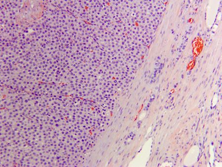
Figure 8. A photomicrograph of a thyroid carcinoma (the patient in this case). Neoplastic epithelial cells are forming dense packets and nests, supported by a fine fibrovascular stroma and surrounded by a thick fibrous capsule (hematoxylin and eosin; 10x).
A high mitotic index is also a common histologic feature of thyroid carcinomas.3 The World Health Organization classifies thyroid carcinomas as well-differentiated, poorly differentiated or undifferentiated.3,4 Well-differentiated thyroid carcinomas are further subdivided into follicular, compact, follicular-compact and papillary carcinomas.1 Follicular-compact and compact are the most common histologic types.1,2,4 There is currently no known association between the histologic subtype and prognosis.1 Regardless of histologic type, thyroid carcinomas often penetrate the capsule, which is considered the primary criterion of malignancy in this tumor type.3 Once the neoplastic cells have escaped the capsule, they will invade the surrounding tissues,2 including the cranial and caudal thyroid veins. Invasion into surrounding veins results in pulmonary metastasis, often prior to regional lymph node metastasis.1,2 Tumor diameter, tumor volume, and bilateral disease have all been associated with distant metastasis.5,6 In addition, gross and histologic evidence of vascular invasion are negative predictors for disease-free survival.4
A possible confounding factor of thyroid follicular carcinoma diagnosis is the potential for compact areas of neoplastic follicular cells to resemble neoplastic C-cells (parafollicular cells). To differentiate thyroid follicular carcinoma from parafollicular tumors, immunohistochemical markers against the respective cell products are employed.1,7 Thyroid follicular carcinomas have positive immunoreactivity for cytoplasmic thyroglobulin,1,7 while parafollicular carcinomas have positive immunoreactivity for cytoplasmic calcitonin.7 Thyroid transcription factor-1 (TTF-1) has also been used as an immunohistochemical marker of thyroid carcinomas; there is some evidence that, when used in conjunction with thyroglobulin or calcitonin, TTF-1 enhances thyroid carcinoma diagnosis.7
References
1. Maxie MG. Endocrine glands. In: Pathology of domestic animals. 6th ed. St. Louis, Missouri: Elsevier; 2016.
2. Meuten DM. Tumors of the endocrine glands. In: Tumors in domestic animals. 4th ed. Ames, Iowa: Iowa State Press;2002.
3. Kiupel M, Capen C, Miller M, et al. Histological classification of tumors of the endocrine system of domestic animals. In: Schulman FY, ed. WHO international histological classification of tumors of domestic animals. Washington, DC: Armed Forces Institute of Pathology, 2008;25-39.
4. Campos M, Ducatelle R, Rutteman G, et al. Clinical, pathologic, and immunohistochemical prognostic factors in dogs with thyroid carcinoma. J Vet Intern Med 2014;28:1805-1813.
5. Thon AP, Marks SL, Feldman ES, et al. Prognostic factors and patterns of treatment failure in dogs with unresectable differentiated thyroid carcinomas treated with megavoltage irradiation. J Am Vet Med Assoc 2000;216:1775-1779.
6. Leav I, Schiller AL, Rijnberk A, et al. Adenomas and carcinomas of the canine and feline thyroid. Am J Pathol 1976;83:61-122.
7. Ramos-Vara JA, Miller MA, Johnson GC, et al. Immunohistochemical detection of thyroid transcription factor-1, thyroglobulin, and calcitonin in canine normal, hyperplastic, and neoplastic thyroid gland. Vet Pathol 2002:39;480-487.