Catch the wave: Urine collection through voiding
This free-flowing form of urine sample collection seems ideal but has a few drawbacks. The main one-possible bacterial contamination. So dont go here if you want to perform a culture.

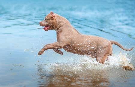

(Photo: Shutterstock.com)Three methods are commonly used for urine collection in canine and feline patients: free-catch, cystocentesis and catheterization.1 When should you opt for free-catch? Read on.
Indications
The advantage of collecting naturally voided urine is that there is little associated patient risk, provided that the bladder is not manually compressed in an excessively forceful manner. Free-catch urine sampling is noninvasive, so obtaining a free-catch urine sample may be beneficial in animals with neoplasia of the urogenital tract, as iatrogenic seeding of the tumor is unlikely to occur. However, when free-catch collection methods are used, urine flows through the distal urethra, genital tract, hair and skin, contaminating the sample with commensal bacterial populations. For this reason, naturally voided urine samples should not be used for urine culture.2 Urine may be collected midstream to decrease concerns of bacterial contamination, but sample contamination remains inevitable.
This collection method is indicated in patients where cystocentesis or catheterization is not indicated or possible. It's useful for evaluating urine specific gravity (USG), semiquantitative chemical analysis and microscopic examination for cells, but it may be associated with artifactual crystalluria and alteration of infectious agents.3 Results of aerobic bacteriological culture are inaccurate using a voided sample due to contamination of distal urogenital and skin organisms.4
What you need
Choosing an appropriate collection container is imperative to preserve sample integrity. Disposable, inexpensive sterile containers with tight-fitting lids help in good sample management and may be provided to the owner prior to collection or purchased from a medical supply store. Transparent containers should be used when available, as they may be helpful in assessing urine color and turbidity.
When an owner obtains a free-catch sample from a dog or cat, the use of improvised containers is discouraged as they may contain contaminants that alter diagnostic testing results. It should be noted that using a sterile collection container does not imply subsequent sample sterility, as it's not a closed system; it simply helps reduce further sample contamination. If samples are not to be immediately submitted for analysis, they should be placed on ice or in a refrigerator for preservation until submission is possible.
How to do it
Dogs. This form of collection is easy to perform in most male dogs as their posture for urination allows ample space for catching urine midstream with a sterile container. When collecting urine from female dogs, a container lid is often easiest. Slide the lid under the dog a few seconds after she begins to urinate, collect 5 to 10 ml (1 to 2 teaspoons), pour the lid contents into the container and close. With this method, in addition to obtaining a urine sample for analysis, you can watch the patient urinate and determine if the process appears normal or abnormal. Don't want to get so close to the source? Sticks and poles are also available to facilitate urine collection while maintaining a distance from the patient.5
Cats. For free-catch collection from male or female cats, one straightforward method is to place the cat in a room for a few hours with an empty litter box or a litter box containing a small amount of nonabsorbable litter. The box should be checked frequently and once urine is noted, it can be poured into a container. Hospitalized patients can be placed in a cage with a raised, nonabsorbable, slatted grate to allow voided urine to pool on the cage floor while keeping the patient clean and dry. The cage should be thoroughly cleaned before placing the patient in the cage to reduce further contamination of the sample, which may interfere with analysis.
Once the urine has been voided, aspirate a sample from the cage floor with an appropriate size syringe (3- to 6-ml). Place the tip of the syringe in the urine, being careful to avoid contacting the floor if possible, and aspirate the urine into the syringe. You can use this same method if a patient has voided urine onto other nonabsorbable surfaces such as an exam table.
If the patient is unwilling to void naturally, you can palpate or gently compress the bladder to encourage urination, but bladder palpation, much less compression, can be difficult to impossible if it contains little or no urine. Compression should never be unduly forceful, as the urinary bladder may be traumatized if excessive pressure is used.
Possible complications
There are minimal complications with collection of voided urine.
References
1. Ackerman N, Aspinall V. Aspinall's complete textbook of veterinary nursing. United Kingdom: Elsevier Health Sciences, 2016.
2. Sorensen TM, Jensen AB, Damborg P, et al. Evaluation of different sampling methods and criteria for diagnosing canine urinary tract infection by quantitative bacterial culture. Vet J 2016;216:168-173.
3. Callens AJ, Bartges JW. Urinalysis. Vet Clin North Am Small Anim Pract 2015;45:621-637.
4. Weese JS, Giguere S, Guardabassi L, et al. ACVIM consensus statement on therapeutic antimicrobial use in animals and antimicrobial resistance. J Vet Intern Med 2015;29:487-498.
5. Vap LM, Shropshire SB. Urine cytology: collection, film preparation, and evaluation. Vet Clin North Am Small Anim Pract 2017;47:135-149.
At the time this article was written, Dr. Morgan Kelley was a veterinary student and Dr. Hilary C. Ludwig was an intern in the Department of Small Animal Medicine and Surgery at the University of Georgia's College of Veterinary Medicine, Athens, Georgia, where Dr. Joseph Bartges is a professor of medicine and nutrition. Dr. Kelley is now a first-year emergency critical care resident at Angell MSPCA and Dr. Ludwig is a resident in small animal surgery at the University of Pennsylvania Ryan Veterinary Hospital, Philadelphia, Pennsylvania. Amanda Callens works at BluePearl Veterinary Partners in Seattle and Renton, Washington. Phillip Snow is the manager and biomedical photographer at the University of Tennessee's College of Veterinary Medicine, Knoxville, Tennessee.

Urinalysis offers a noninasive, rapid screening for canine cancer detection
February 9th 2024This is the first rapid test using urine developed by the Virginia Tech College of Engineering, College of Agriculture and Life Sciences, and the Virginia-Maryland College of Veterinary Medicine
Read More


Sweet pee new remedy in feline diabetes
November 9th 2023A novel class of drugs normalizes blood glucose in type 2 diabetic cats by dumping sugar into urine rather than modulating glucose uptake in the tissues but patient selection and close monitoring are crucial to using them safely
Read More

