Challenging cases in internal medicine: Managing a dog with chronic pruritus and proteinuria
A 14-year-old, 41-lb (18.5-kg), spayed female collie-mix presented for evaluation of intense pruritus and gradually worsening skin disease of more than one year's duration.
A 14-YEAR-OLD, 41-lb (18.5-kg), spayed female collie-mix presented to the Veterinary Medical Teaching Hospital at the University of Wisconsin-Madison for evaluation of intense pruritus and gradually worsening skin disease of more than one year's duration. The skin lesions had begun on the face, ventrum, and feet as diffuse erythema and pruritus. The dog had a history of seasonal pruritus in the fall, and, based on that history, seasonal atopic dermatitis had been presumptively diagnosed. The dog had been treated symptomatically and effectively with oral glucocorticoids and essential fatty acids. But more recently, the pruritus had become unresponsive to therapy, including various combinations of antibiotics, ketoconazole, glucocorticoids, antihistamines, and shampoos. As the pruritus worsened, the dog received increasing doses of glucocorticoids. Unfortunately, the lesions progressed, and the dog's skin was now very pruritic and painful.
In addition to the skin disease, the owner reported that the dog had exhibited polyuria and polydipsia (PU/PD) since it was 11.5 years of age. The PU/PD had been extensively investigated two years earlier at a referral hospital, and pertinent findings included isosthenuria (urine specific gravity = 1.009), a low normal serum albumin concentration (2.5 g/dl; normal = 2.5 to 4 g/dl), and a small nodule on the spleen. At that time, the owner had declined further evaluation of the PU/PD but had authorized an ovariohysterectomy and splenic mass removal. Histopathologic examination of the splenic nodule had revealed benign hyperplasia. The PU/PD had continued over the past two years, and a month before referral, the dog's serum albumin concentration was subnormal at 2 g/dl.
The owner also reported that the dog was depressed, had not been eating or drinking regularly, and was losing weight. Upon further questioning, we discovered the dog would eat if food was presented to it, but it was unwilling to walk to its food bowl, presumably because of its painful footpads.
The owner had acquired the dog when it was 8 months old. The dog's vaccination status was current. The dog had been regularly tested for heartworm disease and had received a preventive monthly.
PHYSICAL EXAMINATION AND INITIAL DIFFERENTIAL DIAGNOSES
On physical examination, the dog was afebrile, depressed, mildly dehydrated, and thin. It was reluctant to walk, and its skin was extremely malodorous. The dog also had severe gingivitis and dental tartar. Ocular and dermatologic lesions included moist periocular erythema and alopecia, mucopurulent conjunctivitis, seborrheic otitis externa, facial excoriations with broken whiskers, lip fold pyoderma, dorsal scaling and thinning of the coat, multifocal areas of deep pyoderma on the dorsum and ventrum characterized by hair matted with blood and pus, and severe pododermatitis. The pododermatitis was characterized by severe interdigital ulceration, erythema, swelling, and exudation. The hair between the footpads was matted with dried exudate and blood, and the footpads were severely crusted and painful when examined. The dog exhibited intense pruritus by rubbing its face and chewing its feet.
Because of the dog's multiple medical problems, we took a practical approach to the differential diagnoses (Table 1). The dog's weight loss and dehydration may have been from the lack of intake (i.e. reluctance to walk to the food and water bowls) or may have resulted from the PU/PD. In an older dog, the most common differential diagnoses for PU/PD and concurrent skin disease include renal disease, diabetes mellitus, hyperadrenocorticism, severe liver disease, hypercalcemia, or a paraneoplastic syndrome. The hypoalbuminemia could have resulted from a protein-losing nephropathy, a protein-losing enteropathy, liver disease, exudative skin disease, vasculitis from an autoimmune disease, or a lack of dietary protein intake.


Table 1: Initial Problems and Differential Diagnoses
With regard to the skin disease, there were two principal issues: the pruritus and presumptive pyoderma. Since pruritus can cause pyoderma and pyoderma can cause pruritus, it is difficult to organize a rational diagnostic plan without first eliminating one of these problems. It is usually best to investigate and eliminate the pyoderma first, and then investigate any residual pruritus. In our experience, the hair loss, scaling, seborrhea, crusting, ulceration, and pruritus were likely to be secondary to severe infection. The most common cause of deep pyoderma in a dog is demodicosis. The term pyoderma literally means pus in the skin, so any disease causing a neutrophilic exudate results in pyoderma. In addition to demodicosis and infectious skin diseases (e.g. intermediate and deep mycoses, nonstaphylococcal infections), we also considered neoplasia. Immune-mediated skin diseases, such as lupus erythematosus, were considered possible. Hepatocutaneous syndrome (i.e. profound dermatologic manifestations secondary to severe liver disease) was considered because of the footpad crusting and hypoalbuminemia, but the generalized deep pyoderma was inconsistent with the classic clinical signs of this syndrome.
DIAGNOSTIC TESTING
Although we recommended concurrent investigation of the skin disease and PU/PD, financial constraints and owner concerns necessitated a workup of the dog's skin disease first. Evaluation of multiple skin scrapings from the dog's feet, face, and trunk revealed large numbers of Demodex mites. Cytologic evaluation of impression smears of skin exudate revealed large numbers of degenerate neutrophils, intracellular and extracellular cocci, and five to 10 Malassezia organisms per high-power field. Large numbers of yeast were also found in ear swabs. The dog's packed cell volume was 42% (normal = 37% to 55%), and its total solids measurement was 8.4 g/dl (normal = 5 to 8 g/dl). The owner declined a complete blood count and serum chemistry profile but authorized urine testing. Urinalysis revealed a specific gravity of 1.035 with 4+ protein and a normal urine sediment. Because of the marked proteinuria and historical hypoalbuminemia, we measured the urine protein:creatinine ratio. The result was 5.35 (normal < 1).
DERMATOLOGIC TREATMENT AND FOLLOW-UP
Initial treatment and diagnoses
Our initial diagnosis was adult-onset demodicosis complicated by a deep bacterial and Malassezia species pyoderma. In addition, the urinary protein loss could explain the low serum albumin concentration detected by the referring veterinarian. The owner declined further diagnostic testing for the proteinuria until the dog showed a response to therapy for the skin disease, which was a quality-of-life issue at the time of presentation.
The dog was hospitalized and treated for pain with butorphanol tartrate (0.5 mg/kg orally b.i.d.). We clipped the dog's hair and administered a warm-water whirlpool treatment and chlorhexidine bath to remove the matting and crusts on the face, feet, and trunk. In addition, we addressed supportive nutritional and fluid therapy needs by hand-feeding a high-calorie palatable soft food, administering subcutaneous fluids (lactated Ringer's solution, 30 ml/kg b.i.d.), and directly offering water by hand several times a day. We initiated cephalexin (30 mg/kg orally b.i.d. for 45 days) for the bacterial pyoderma, and itraconazole (5 mg/kg orally once a day for 10 days) for the Malassezia species infection. We chose itraconazole because it is less likely than ketoconazole to cause side effects, including a decreased appetite. The extensive deep pyoderma precluded treatment with amitraz for the demodicosis, so we administered milbemycin oxime (3 mg/kg orally once a day for 30 days pending reevaluation). Amitraz was also avoided because it can cause insulin resistance with subsequent glucosuria and PU/PD. Ivermectin was not used because the dog appeared to be a collie-mix.
Three days after presentation, the dog was discharged from the hospital. The owner was instructed to continue the oral medications and to bathe the dog with chlorhexidine shampoo at least every other day until the skin exudation and hair matting had resolved. Although the dog's pruritus was markedly decreased at discharge, the owner was concerned about the dog's comfort, so we prescribed fexofenadine hydrochloride (Allegra—Aventis; 2 mg/kg orally b.i.d.) until the next recheck examination. At presentation, the dog had been receiving prednisone (0.5 mg/kg orally once or twice a day). After much consideration about whether to taper or simply discontinue this anti-inflammatory dosage of prednisone, we stopped it without tapering. Glucocorticoid use in a patient with severe deep pyoderma, demodicosis, and PU/PD is contraindicated, and we strongly felt it was a contributing factor in the development of adult-onset demodicosis in this dog. During hospitalization, an unquestionably stressful event, the dog became more active, began to eat, and showed no signs of glucocorticoid withdrawal.
Follow-up
During a follow-up examination two weeks later, the owner reported that the dog was much brighter and was walking normally, even going for walks. The dog's skin and paws were markedly improved, with minimal pruritus, evidence of coat regrowth, and resolving deep pyoderma, although superficial pyoderma was still present. The dog's skin was mildly erythematous and somewhat oily. Evaluation of impression smears revealed no Malassezia organisms. Cytologic examination of skin scrapings revealed large numbers of adult Demodex mites, which were all dead. The only other dermatologic abnormality noted was easily bruised, thin skin. There were larger-than-expected petechiae, but only in the areas where we performed skin scrapes. Easy bruising after skin scraping is common in dogs with inflamed skin, especially in those receiving glucocorticoids. We continued oral cephalexin and milbemycin.
At a recheck one month later, the dog's skin was almost normal except for residual hyperpigmentation and mild seborrheic otitis. Skin scraping results remained positive, but again only dead adult Demodex mites were found. At this time, the owner was optimistic about the dog's recovery and was now willing to investigate the proteinuria and hypoalbuminemia, so we had the internal medicine service evaluate the dog (see below).
Long-term dermatologic management
During the next four months, the dog's bacterial pyoderma and pododermatitis resolved. Six weeks and 10 weeks after starting milbemycin therapy, the dog's skin scraping results were negative for Demodex species. Milbemycin therapy was continued for about 14 weeks. Skin scrapings were performed every two or three months, and the dog experienced a relapse of demodicosis eight months after its initial presentation. We restarted milbemycin at the previous dosage. This resolved the dog's clinical signs, but skin scraping results remained positive for Demodex mites, so lifelong milbemycin therapy was recommended.
EVALUATION AND TREATMENT OF INTERNAL MEDICINE PROBLEMS
About a month after the adult-onset demodicosis and deep pyoderma were diagnosed, the dog presented to the internal medicine service for evaluation of proteinuria and hypoalbuminemia. According to the owner, the dog's overall attitude greatly improved as the pyoderma resolved, but its appetite was still decreased. The polydipsia remained, but the dog was urinating normally. Physical examination revealed a quiet to depressed dog. Its hydration status, temperature, and pulse and respiratory rates were normal. There was a generalized thinning of the coat, with evidence of hair regrowth, patchy erythema, and multifocal crusting. The dog had severe dental tartar, gingivitis, and presumed periodontitis. The dog was somewhat thin, although its body weight (41 lb) was basically unchanged from the initial evaluation by the dermatology service.
The results of the urinalysis performed at presentation a month earlier ruled out diabetes mellitus, renal insufficiency, and diabetes insipidus as causes of the PU/PD. There was no glucosuria, and a urine specific gravity of 1.035 in a dehydrated dog was evidence that the kidneys were capable of producing concentrated urine. Other differential diagnoses for the dog's PU/PD included iatrogenic or spontaneous hyperadrenocorticism, occult pyelonephritis, hypercalcemia, psychogenic polydipsia, and liver disease. The differential diagnoses for the proteinuria and historic hypoalbuminemia included glomerulonephritis, amyloidosis, pyelonephritis, and chronic interstitial nephritis. The urine protein:creatinine ratio of 5.35 was greater than expected for chronic interstitial nephritis and less than expected for renal amyloidosis.1 Because granular casts, white blood cells, and red blood cells were not present in the urine sediment, pyelonephritis was unlikely. These observations, along with the history of chronic inflammatory skin disease, made glomerulonephritis the most likely differential diagnosis. Glucocorticoid use can lead to proteinuria,2 but considering the historical hypoalbuminemia in this dog, it was more likely that glucocorticoid use either predisposed the dog to glomerulonephritis or exacerbated preexisting glomerulonephritis.3
Diagnostic tests
A complete blood count, serum chemistry profile, and urinalysis were done. A urine bacterial culture and antimicrobial sensitivity testing were performed after cystocentesis to help rule out pyelonephritis. The urine protein:creatinine ratio was reevaluated to see if it had improved as a result of therapy for the skin diseases and cessation of exogenous glucocorticoids. A thoracic radiographic examination was performed to screen for neoplasia, abscesses, or chronic granulomatous diseases. An abdominal ultrasonographic examination was performed to search for neoplasia, organomegaly, adrenal gland pathology, and urolithiasis, as well as to evaluate renal architecture. The dog's systolic blood pressure was measured with a Doppler unit, because dogs with glomerulonephritis have a high incidence of systemic hypertension.4 The results of the diagnostic tests were normal, with a few exceptions. The urine protein:creatinine ratio had markedly improved to 2.22. The urine specific gravity was 1.030, with 3+ protein and a normal urine sediment. The serum albumin concentration was still low (2.1 g/dl), with normal concentrations of globulin (3.6 g/dl; normal = 2.6 to 4.4 g/dl) and total protein (5.7 g/dl). The cholesterol concentration was elevated (344 mg/dl; normal = 111 to 290 mg/dl). The dog's systolic blood pressure was normal at 130 mm Hg. Seven readings were recorded; the highest and lowest were excluded, and the remaining five were averaged. Further diagnostic testing was recommended for the following week.
An ACTH stimulation test was performed to rule out hyperadrenocorticism, and a bile acid tolerance test was performed to rule out hepatic dysfunction as possible causes for the PU/PD. The dog had not received glucocorticoids for about a month, which is an adequate time to allow for a meaningful interpretation of the ACTH stimulation test. Because infectious diseases are known to cause glomerulonephritis, serologic testing for infectious diseases common to the dog's geographic area (e.g. ehrlichiosis, leptospirosis, Lyme disease) was performed. The findings from these tests were normal.
A thyroid panel was performed to rule out concurrent hypothyroidism as a cause for the dog's continued depression and dermatologic disease. Although this could have been performed on initial evaluation by the dermatology service, interpretation of thyroid testing at that time would have been more difficult because of nonthyroidal illness and glucocorticoid administration.
The thyroid panel revealed a slightly low total T4 concentration (13 nmol/L; normal = 15 to 67 nmol/L), a normal free T4 concentration by dialysis (18 pmol/L; normal = 6 to 42 pmol/L), and an elevated canine TSH concentration (77 mU/L; normal = 0 to 37 mU/L). There was no increase in antibodies against T4, T3, or thyroglobulin. These discordant findings were consistent with sick euthyroid syndrome.5 Early hypothyroidism or a spuriously normal free T4 result was another possibility. Repeat thyroid testing over time was advised, particularly if the skin disease did not respond to appropriate therapy.
Treatment and follow-up
Because the urine protein:creatinine ratio had improved and the owner was reluctant to allow invasive testing, a renal biopsy was not performed. Without histopathologic examination of a renal biopsy, we could not provide a definitive diagnosis for the cause of the dog's proteinuria. The decrease in urine protein loss may have resulted from cessation of exogenous glucocorticoids and improvement in the inflammatory skin disease. Because the dog continued to have proteinuria (i.e. urine protein:creatinine ratio > 1) and the hypoalbuminemia had not resolved, we prescribed an angiotensin-converting enzyme inhibitor (enalapril maleate, 0.5 mg/kg orally once a day) to further reduce the proteinuria and perhaps slow the progression of chronic renal disease.6 Low-dose aspirin therapy (0.5 mg/kg orally once a day) was prescribed to ameliorate platelet-mediated glomerular inflammation and to reduce the thromboembolic tendency often associated with protein-losing renal disease.6,7 The referring veterinarian later prescribed carprofen for degenerative joint disease, so aspirin therapy was discontinued because of possible side effects when two nonsteroidal anti-inflammatory drugs are used. The dog was eating a premium-quality senior food, moderately restricted in protein, which was considered appropriate for this stage of the dog's renal disease.
Laboratory testing performed before the much-needed dentistry showed the dog's serum albumin concentration had increased to 2.5 g/dl and the blood urea nitrogen and creatinine concentrations remained normal. A urinalysis revealed 1+ protein, a specific gravity of 1.016, and a normal sediment. Table 2 summarizes urine testing results over time in this patient as they relate to the status of the skin disease. Although the proteinuria improved, it never resolved. It is not possible to assess the relative contribution of the enalapril vs. other aspects of the case management (e.g. treatment of demodicosis and bacterial pyoderma, cessation of glucocorticoid therapy) to the improvement in the proteinuria.

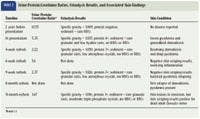
Table 2: Urine Protein:Creatinine Ratios, Urinalysis Results, and Associated Skin Findings
DISCUSSION
Adult-onset canine demodicosis presents in one of two patient populations. The first is a group of dogs with a history of demodicosis as a puppy or young adult. The disease either spontaneously resolves or is, apparently, successfully treated. These dogs remain clinically normal until something triggers a relapse later in life. It is speculated that these dogs develop initial disease because of an inherent defect in the skin's immune system. The second is a group of dogs with no known history of demodicosis as a puppy or young adult that develop disease for the first time as older adults (i.e. usually over 5 years of age).8
In this second group of dogs, or dogs with true adult-onset demodicosis, it is unclear what triggers the disease. Obviously, the host has been living in harmony with its Demodex mite population for years until something changes. The most commonly cited cause is an underlying medical condition that debilitates or immunocompromises the host (e.g. diabetes mellitus, hyperadrenocorticism, hypoadrenocorticism, excessive glucocorticoid administration, hypothyroidism, neoplasia). In many dogs, the underlying disease may not be apparent when the demodicosis is diagnosed. One of the authors (Dr. Moriello) has observed adult-onset demodicosis to precede the detection of a perianal adenocarcinoma by one year. That dog's demodicosis was unresponsive to medical treatment but resolved with surgical excision of the tumor a year later when it was discovered.
The diagnosis and treatment of adult-onset demodicosis is a two-step process. First, the demodicosis needs to be diagnosed, usually by skin scrapings. Multiple deep skin scrapings (> 5) are recommended. The most common cause of missing the diagnosis is to do too few skin scrapings. Although Demodex mites are a normal part of the skin's fauna, they are rarely found in skin scrapings from healthy dogs. So their presence on a skin scraping should alert you to a problem.8 Second, the underlying trigger for the adult-onset demodicosis needs to be investigated. This investigation can be difficult, expensive, and sometimes unrewarding.
Treatment of adult-onset demodicosis is similar to that of juvenile demodicosis. Concurrent bacterial and yeast pyodermas should be treated for at least 30 days. If a deep pyoderma is present, delay topical therapy until it is healed. Clip the hair for easier medicated bathing (select a follicular flushing or an antimicrobial shampoo based on patient assessment) and, if and when appropriate, administer weekly amitraz dips. It is unclear which of the various treatments are most effective in dogs with generalized demodicosis, particularly adult-onset forms. A critical factor in treatment success is whether the underlying trigger can be identified and cured or at least managed. If not, then it is unlikely the demodicosis will be cured, and the dog's clinical signs will only be managed. Regardless of the treatment protocol, continue treatment for at least 30 days past mite cure (i.e. negative results for two skin scrapings at weekly or biweekly intervals). Treatment considerations for demodicosis are listed in Table 3.

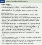
Table 3: Treatment Considerations for Demodicosis
Investigating the underlying cause of adult-onset demodicosis requires obtaining a thorough history. One of the authors (Dr. Moriello) had a canine patient whose adult-onset demodicosis was secondary to its owner's excessive application of a human prescription betamethasone ointment to the dog's skin. In other cases, the cause is more obvious (e.g. the dog has hypothyroidism or diabetes mellitus, or it has been receiving exogenous oral or injectable glucocorticoids). Dogs with adult-onset demodicosis may require extensive evaluation to identify the underlying cause of the mite overpopulation. This evaluation may include a complete blood count, a serum chemistry profile, urinalysis, urine cultures, thoracic and abdominal radiography and ultrasonography, ACTH stimulation testing, low-dose dexamethasone suppression testing, and a thyroid profile.
In this case, it was difficult to pinpoint what triggered and perpetuated the dog's adult-onset demodicosis. The dog may have had its usual episode of seasonal atopic dermatitis the previous fall and then developed a concurrent bacterial and yeast pyoderma. Inappropriate therapy may have led to a lack of clinical response, and glucocorticoid use may have triggered the adult-onset demodicosis. Because the dog had been acquired at 8 months of age, juvenile demodicosis cannot be ruled out. Also, the low albumin and elevated cholesterol concentrations and proteinuria must be considered. The skin requires a great deal of protein each day for homeostasis, and any protein loss from the body or decrease in protein production or daily intake will be reflected in the health of the skin.
A definitive cause for the chronic PU/PD was not ascertained. The finding of relatively concentrated urine (specific gravities of 1.035 and 1.026) on two separate occasions made the observation of PU/PD by the owner suspect; the problem may have been intermittent or associated with glucocorticoid use.
Although a histologic diagnosis of the kidney disease was not obtained, glomerulonephritis was tentatively diagnosed. The degree of elevation of the urine protein:creatinine ratio, probable exclusion of pyelonephritis (based on results of the urinalysis, urine culture, and renal ultrasonographic examination), and the finding of hypercholesterolemia are all consistent with glomerulonephritis. Glomerulonephritis can be idiopathic or result from infectious, inflammatory, or neoplastic disease. Although the only infectious diseases identified in this case were the periodontitis and the pyoderma, the chronicity and severity of these inflammatory processes could have been a source for antigen:antibody complexes that could result in secondary glomerulonephritis. Dogs with generalized demodicosis and recurrent staphylococcal pyoderma have been reported to have significantly higher mean circulating immune complexes compared with normal dogs.9 At least one breed of dog, the soft-coated wheaten terrier, which has a high incidence of protein-losing nephropathy, has also been noted to have an increased incidence of inflammatory skin disease (e.g. atopy, pyoderma), although the cause-and-effect relationship is unknown.10 Alternatively, a more global disorder of the immune system could have resulted in both the adult-onset demodicosis and the presumptive glomerulonephritis. Since a renal biopsy was not obtained, it is also possible that this dog had chronic interstitial nephritis of unknown cause with an unusual degree of proteinuria. But it would be unusual to find adequately concentrated urine in a dog with marked chronic interstitial nephritis (i.e. severe enough to cause proteinuria, hypoalbuminemia, and hypercholesterolemia). Whatever the renal diagnosis, the proteinuria in this case was exacerbated by glucocorticoid use or the deep pyoderma, since the proteinuria improved markedly with glucocorticoid discontinuation and appropriate treatment of the skin diseases, even before treatment with enalapril.
It is interesting to relate the urine protein:creatinine ratios to the presence or absence of skin disease (Table 2). The urine protein:creatinine ratio was at its highest when the dog was most symptomatic (i.e. untreated severe deep pyoderma and demodicosis). At this point the dog's prognosis seemed poor, and severe, irreversible, and worsening renal disease as an underlying trigger of the adult-onset demodicosis was discussed with the owner. It was surprising that the urine protein:creatinine ratio markedly improved and almost reached normal when the dog's skin disease was in remission. Interestingly, the urine protein:creatinine ratio worsened when the dog's skin disease relapsed. Clearly, it seems concurrent disease can affect this renal function test. An important lesson in this case is that decisions about treatment, prognosis, and, in particular, euthanasia, should be made only after obtaining several urine protein:creatinine ratio measurements, preferably after treating concurrent disease.
Adult-onset demodicosis and protein-losing kidney disease are serious disorders that carry poor prognoses. Fortunately, this dog responded well to therapy for both conditions, and the owner eventually allowed dentistry to address the periodontal disease. The improved quality of life certainly extended this dog's life, because the owners had been considering euthanasia because of the dog's discomfort from its skin problems. Alternatively, if the protein-losing renal disease had been allowed to progress, as it usually does without intervention, the dog might have succumbed to renal failure or a complication of the hypoproteinemia, such as a thromboembolic event.
Karen A. Moriello, DVM, DACVD
Melissa S. Wallace, DVM, DACVIM
Department of Medical Sciences
School of Veterinary Medicine
University of Wisconsin
Madison, WI 53706
REFERENCES
1. Center, S.A. et al.: 24-Hour urine protein/creatinine ratio in dogs with protein-losing nephropathies. JAVMA 187 (8):820-824; 1985.
2. Waters, C.B. et al.: Effects of glucocorticoid therapy on urine protein-to-creatinine ratios and renal morphology in dogs. J. Vet. Intern. Med. 11 (3):172-177; 1997.
3. Center, S.A. et al.: Clinicopathologic, renal immunofluorescent, and light microscopic features of glomerulonephritis in the dog: 41 cases (1975-1985). JAVMA 190 (1):81-90; 1987.
4. Cook, A.K.; Cowgill, L.D.: Clinical and pathological features of protein-losing glomerular disease in the dog: A review of 137 cases (1985-1992). JAAHA 32 (4):313-322; 1996.
5. Peterson, M.E. et al.: Measurement of serum total thyroxine, triiodothyronine, free thyroxine, and thyrotropin concentrations for diagnosis of hypothyroidism in dogs. JAVMA 211 (11):1396-1402; 1997.
6. Grauer, G.F. et al.: Effects of enalapril versus placebo as a treatment for canine idiopathic glomerulonephritis. J. Vet. Intern. Med. 14 (5):526-533; 2000.
7. Grauer, G.F.: Glomerulonephritis. Semin. Vet. Med. Surg. 7:187-197; 1992.
8. Scott, D.W. et al.: Canine demodicosis. Muller and Kirk's Small Animal Dermatology, 6th Ed. W.B. Saunders, Philadelphia, Pa., 2000; pp 457-476.
9. DeBoer, D.J. et al.: Circulating immune complex concentrations in selected cases of skin disease in dogs. AJVR 49 (2):143-146; 1988.
10. Littman, M.P. et al.: Familial protein-losing enteropathy and protein-losing nephropathy in Soft Coated Wheaten Terriers: 222 cases (1983-1997). J. Vet. Intern. Med. 14 (1):68-80; 2000.