Clinical Exposures: Canine circumanal gland adenoma: The cytologic clues
A 9-year-old spayed female golden retriever was presented to the Tufts Veterinary Emergency Treatment and Specialties hospital for evaluation of a perianal mass.
A 9-year-old spayed female golden retriever was presented to the Tufts Veterinary Emergency Treatment and Specialties hospital for evaluation of a perianal mass. The owners had first noticed the mass a few weeks earlier, and no change in the lesion’s size, shape, or character had occurred since that time. The mass appeared to be nonpainful to the patient. At presentation, the mass was 1.5 cm in diameter and about 0.5 cm ventral to the anus. Grossly, the subcutaneous lesion appeared smooth, firm, and nonulcerated. The remainder of the patient’s physical examination was unremarkable. Fine-needle aspiration biopsy of the perianal mass was performed.
CYTOLOGIC EXAMINATION FINDINGS
Cytologic evaluation of the aspirate revealed a prominent population of round-to-polygonal nucleated cells arranged as cohesive groups with isolated individual cells (Figure 1A).
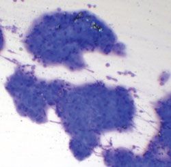
1A. Cytologic examination of a fine-needle aspirate from the perianal tumor of the patient in this case report. Several clusters of tightly adherent round-to-polygonal epithelial cells can be seen (Wright’s-Giemsa stain; 100X).
These cells had abundant finely granular, amphophilic cytoplasm. The nuclei were round-to-oval with reticular chromatin and one to three prominent nucleoli (Figure 1B). A mild degree of anisocytosis and anisokaryosis was observed. In addition, smaller reserve type cells, with darker cytoplasm and a higher nucleocytoplasmic ratio, were present along the margins of some clusters. A small number of erythrocytes and occasional nondegenerate neutrophils and large mononuclear cells (i.e. monocytes or macrophages) were noted in the background. No infectious organisms were found.
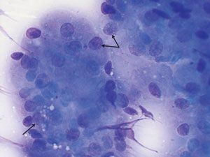
1B. At high power, the finely granular and often amphophilic cytoplasm can be appreciated. Note the single, prominent nucleolus (black arrows) in most of the individual cells (Wright’s-Giemsa stain; 1,000X).
These cytologic findings were consistent with a well-differentiated circumanal gland tumor, also referred to as perianal tumor or hepatoid gland tumor. Given the relatively uniform appearance (i.e. lack of cellular or nuclear atypia observed) of the epithelial cells, it was presumed likely that this tumor was a benign adenoma. However, excision and histopathologic evaluation of tumor margins for tissue or vascular invasion is often necessary to identify a circumanal gland carcinoma.
TREATMENT AND OUTCOME
The perianal tumor was surgically removed. Histologic evaluation of the biopsied tissue verified the cytologic diagnosis and confirmed that the circumanal gland adenoma was completely excised. The patient did well postoperatively.
About one year later, this patient was referred to Tufts for an unrelated emergency. At that time, no evidence of regrowth or recurrence of the circumanal gland adenoma was noted on physical examination.
DISCUSSION
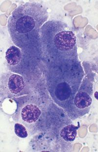
Circumanal glands are modified sebaceous glands unique to the Canidae family (coyotes, dogs, foxes, jackals, and wolves)1-4 and are typically located in the perianal region. These glands are also found in areas such as the perineum, dorsal and ventral tail base, upper thigh and hindlimbs, and dorsal midline and lumbar regions, in addition to the preputial area in male dogs and the abdominal mammary region in female dogs.1-3 The individual epithelial cells that make up the circumanal glands are cytologically and histologically similar in appearance to hepatocytes (Figure 2A & 2B). Both cell types have abundant, prominently granular cytoplasm and round nuclei with a prominent nucleus. So circumanal glands and circumanal gland adenomas may also be referred to as hepatoid glands and hepatoid gland adenomas, respectively.1-3
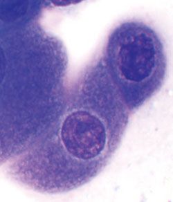
2A. Cytologic examination of a fine-needle aspirate from a circumanal gland adenoma in a different dog illustrating the characteristic polygonal shape and prominent cytoplasmic granularity (Wright’s-Giemsa stain; 1,000X).
2B. Cytologic examination of a fine-needle aspirate from a canine liver showing the cytologic features of normal canine hepatocytes. Note the similarity in morphologic appearance between cells of a circumanal gland adenoma and hepatocytes, leading to the common alternative term for these perianal tumors, hepatoid tumors (Wright’s-Giemsa stain;1,000X).
Circumanal gland adenomas are common tumors in dogs, comprising about 8% to 9% of all epithelial tumors.2,3,5 Most circumanal gland adenomas (about 90%) are found in the perianal region, and these tumors account for more than 80% of all perianal tumors in dogs.1-3 However, circumanal gland adenomas can also arise in the other locations in which the glands can be found. Because the development of these adenomas is largely dependent on the influence of testosterone and other androgens on the circumanal glands, the tumors are classically observed in middle-aged to older intact male dogs. However, circumanal gland adenomas also occur in male neutered dogs (about 20%), intact and spayed female dogs (about 9% and 19%, respectively),6 and, occasionally, young adults (possibly as young as 2 years of age) of either sex.1-3,5
These tumors are typically nodular but may vary in size and diameter. The tumors may present as a single mass or as multiple masses. Depending on where the tumor is located, the masses may be variably raised, ulcerated, or hairless.1-4 Several dog breeds are reported to have an increased predisposition to developing circumanal gland tumors, including Siberian huskies, Samoyeds, Pekingese, and cocker spaniels.1-4
In spite of their relative cytologic pleomorphism (i.e. anisocytosis and multiple nucleoli), circumanal gland adenomas are considered benign or low-grade neoplasms.1,4 Malignant variants (epitheliomas and carcinomas) are uncommon but exhibit a similar pattern of anatomical location and sex predisposition as the benign adenoma. The malignant variants can often be difficult to distinguish from a benign adenoma based on the morphologic features of the individual cells unless the severe cytologic atypia is present. Circumanal gland epitheliomas (low-grade malignancy) are composed primarily of reserve cells and show disorderly growth. Circumanal gland carcinomas are composed of undifferentiated, pleomorphic cells and will show invasion into the surrounding connective tissue and into lymphatics. The latter findings are the most consistent signs of a malignant neoplasm, so histological evaluation of tumor margins and examination of regional lymph nodes are necessary for a definitive diagnosis.2,3
Differential diagnoses
The primary differential diagnosis for a circumanal gland adenoma located in the perianal region is an anal sac adenocarcinoma (arising from the apocrine glands of the anal sac). Cytologically, this malignant tumor looks much different than a circumanal gland adenoma looks. True to the epithelial cell origin of anal sac adenocarcinoma, the round-to-cuboidal tumor cells often cluster, but in a much less cohesive manner than those of circumanal gland tumors, and cell margins are less distinct within these cell aggregates (Figure 3A).
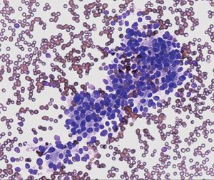
3A. Cytologic examination of a fine-needle aspirate from an anal sac adenocarcinoma in a dog. Note the lack of distinct cellular margins within the aggregations of neoplastic cells (Wright’s-Giemsa stain; 200X). (Photomicrograph courtesy of Cheryl Stockman, MT [American Society for Clinical Pathology].)
Close apposition of nuclei within cell clusters reflects the fact that these cells have only a small amount of basophilic cytoplasm. The neoplastic cells of an anal sac adenocarcinoma typically have a somewhat uniform appearance, and little evidence of cellular or nuclear atypia is present (Figure 3B), suggesting a relatively low-grade or benign tumor. However, these tumors are known to be aggressively infiltrative, often metastasizing locally (i.e. regional sublumbar lymph nodes), and may spread to distant tissue sites such as the liver, spleen, lungs, and vertebral spine.4,7 About 26% to 53% of dogs with anal sac adenocarcinoma have humoral hypercalcemia of malignancy, which can itself be life-threatening.4,7
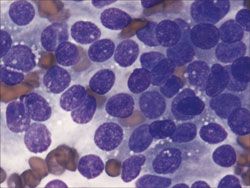
3B. Cytologic examination of a fine-needle aspirate from an anal sac adenocarcinoma in a dog. The relatively uniform and homogeneous appearance of the neoplastic cells may be observed. There is little to no evidence of cellular or nuclear atypia (i.e. criteria of malignancy) (Wright’s-Giemsa stain; 1,000X). (Photomicrograph courtesy of Cheryl Stockman, MT [ASCP].)
If a secondary infection, hemorrhage, or necrotic tissue is present within a perianal mass, or if the sample cellularity is very low or poorly preserved, it may be difficult to cytologically distinguish a circumanal gland adenoma from an anal sac adenocarcinoma.4 Biopsy and histologic evaluation may then be necessary for a definitive diagnosis.
Other differential diagnoses for perianal masses include a plasmacytoma, squamous cell carcinoma, basal cell neoplasia, or skin adnexal tumors. The distinctive granular cytoplasm of the circumanal gland adenoma cells can be used to distinguish this tumor type from these other tumor types.
Therapy and prognosis
Complete excision of circumanal gland adenomas should be curative, but additional tumors may develop from nearby hyperplastic circumanal glands, giving the illusion of recurrence. In intact male dogs, castration alone may cause tumor regression, but neutering with tumor removal (or other adjuvant therapy) is routinely recommended.1,4 In cases of recurrent tumors not amenable to further surgical intervention, adjuvant radiation therapy may be warranted, having a 69% chance of complete tumor control over a two-year period.2-4,7
Alternatively, if surgery is not an option, cryosurgery or estrogen therapy (e.g. diethylstilbestrol) may be considered, but these modalities may be undesirable because of possible complications. Cryotherapy is most effective against focal tumors less than 1 to 2 cm in diameter.4 It is not recommended in the case of large or diffusely infiltrative tumors, or, alternatively, if the tumor is close to the anus, cryosurgery may damage the anal sphincter, resulting in dyschezia or fecal incontinence.2-4 In addition, estrogen therapy is associated with potentially irreversible bone marrow suppression (aplastic pancytopenia) or, in male dogs, squamous metaplasia of the prostate.1,2,4
Conclusion
Circumanal gland adenomas are a common tumor in dogs. The tumor’s location and distinctive cytologic appearance are often sufficient for a cytologic diagnosis. The ability to cytologically diagnose a circumanal gland adenoma is critical in order to differentiate this benign perianal tumor from anal sac adenocarcinoma, which characteristically has significant malignant potential in dogs.
This case report was provided by Maria Vandis, DVM, and Joyce S. Knoll, VMD, PhD, DACVP, Department of Biomedical Sciences, Cummings School of Veterinary Medicine, Tufts University, North Grafton, MA 01536.
REFERENCES
1. Goldschmidt MH, Hendrick MJ. Tumors of the skin and soft tissues. In: Tumors in domestic animals. 4th ed. Ames: Iowa State Press, 2002;68-73.
2. Goldschmidt MH, Shofer FS. Hepatoid gland tumors. In: Skin tumors of the dog and cat. Oxford, U.K.: Pergamon Press, 1992;66-74.
3. Goldschmidt MH, Shofer FS. Anal sac gland tumors. In: Skin tumors of the dog and cat. Oxford, U.K.: Pergamon Press, 1992;105-108.
4. Turek MM, Withrow SJ. Tumors of gastrointestinal tract, H. Perianal tumors. In: Small animal clinical oncology. 4th ed. St. Louis, Mo: Saunders Elsevier, 2007; 503-510.
5. Raskin RE. Skin and subcutaneous tissues. In: Atlas of canine and feline cytology. Philadelphia, Pa.: W.B. Saunders Company, 2001;65-74.
6. Goldschmidt MH, McManus P, Goldschmidt K. Hepatoid gland adenoma. University of Pennsylvania School of Veterinary Medicine's Computer Aided Learning program, 2000. Available at: http://cal.vet.upenn.edu/projects/derm/Home/ADNEXAL/sebac/hepgad.htm.
7. Williams LE, Gilatto JM, Dodge RK, et al. Carcinoma of the apocrine glands of the anal sac in dogs: 113 cases (1985-1995). J Am Vet Med Assoc 2003;223(6):825-831.
Urinalysis offers a noninasive, rapid screening for canine cancer detection
February 9th 2024This is the first rapid test using urine developed by the Virginia Tech College of Engineering, College of Agriculture and Life Sciences, and the Virginia-Maryland College of Veterinary Medicine
Read More