Clinical Exposures: Canine dermatophyte infection
A 3-month-old intact male boxer was presented for a preadoption evaluation at the Foster Hospital for Small Animals at Tufts University after being rescued in North Carolina.
A 3-month-old intact male boxer was presented for a preadoption evaluation at the Foster Hospital for Small Animals at Tufts University after being rescued in North Carolina. The physical examination results were unremarkable except for mild, patchy alopecia on the frontal aspect of the head and a 3-x-2-cm dermal, nodular lesion on the left lateral thigh. These skin lesions were nonpruritic and nonulcerated.
CYTOLOGIC EXAMINATION FINDINGS
Demodex and Sarcoptes species mites were not found on microscopic examination of samples obtained by deep skin scraping of the alopecic area on the head. Cytologic examination of the skin scraping revealed low numbers of erythrocytes, multiple broken hair shafts and anucleate squamous epithelial cells, and small to moderate amounts of keratin debris.
Samples from the thigh lesion were obtained by fine-needle aspiration, and the slides were stained with Wright's-Giemsa. Cytologic examination revealed moderate numbers of erythrocytes and intact nucleated cells. Moderate numbers of inflammatory cells were found, with a predominance of nondegenerate neutrophils and lesser numbers of activated macrophages. A few small lymphocytes and eosinophils were also noted. Some macrophages contained phagocytosed cellular debris and rare hematoidin crystals.
The samples from the thigh lesion also contained a small population of superficial squamous epithelial cells—both individual cells and variably sized aggregates—mixed with varying amounts of keratin debris. A few squamous epithelial cells contained cytoplasmic melanin granules, and others had multiple adherent bacteria.
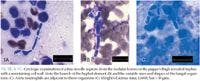
Fragmented fungal hyphae and small, solitary, irregularly shaped structures were also identified in the samples (Figure 1A), all with a basophilic interior and a prominent nonstaining cell wall. The hyphae were septate and exhibited occasional branching (45 to 60 degrees) (Figure 1B). The solitary structures were about 3-μm-x-3-to-5-μm. Most of the structures were round to oval, but some were cuboidal or cigar-shaped or hand-mirror-shaped with a clubbed end (Figure 1C). These structures appeared to be either detached globoid ends of hyphae or spores.


DIFFERENTIAL DIAGNOSES
The presumptive diagnosis was mixed inflammation secondary to infection with a nonpigmented fungus. Differential diagnoses for mycotic infections that form hyphal elements in tissues include dermatophytosis (e.g.Microsporum species, Trichophyton mentagrophytes) and infection with Aspergillus or Penicillium species or other nonpigmented fungi (e.g. Acremonium, Fusarium, Geotrichum, Paecilomyces, Pseudallescheria, and Scedosporium species). Zygomycosis (e.g. Basidiobolus and Conidiobolus species) and pythiosis were also considered because of the puppy's geographic origin.1
HISTOLOGIC EXAMINATION AND DEFINITIVE DIAGNOSIS
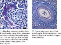
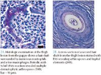
Soon after the preadoption examination, the puppy was neutered, and the thigh lesion was excised with wide margins for histologic examination. The histologic examination findings included a multifocal mixed inflammatory cell (primarily epithelioid macrophages and fewer neutrophils) infiltration of the dermis centered on the hair follicles. Moderate hyperplasia and hyperkeratosis were noted in the epidermis, consistent with self-trauma. Additionally, fungal arthrospores and hyphae were found within the hair shafts and within foci of inflammation (Figures 2A & 2B).

Granulomatous folliculitis and furunculosis with intralesional dermatophyte arthrospores and hyphae were diagnosed. The fungal elements were consistent with dermatophytosis, specifically Microsporum canis infection. The nodular form of dermatophytosis was suspected, but fungal culture to confirm the genus and species of the dermatophyte was not done.
RECOVERY AND FOLLOW-UP
The puppy had an uneventful recovery from surgery. No other treatment was given for the dermatophytosis. The lesions on the head resolved over several weeks without intervention. Eight months later, the puppy was in a new home and doing well. No new skin lesions had developed.
DISCUSSION
Dermatophytosis, also called ringworm, may be caused by several keratinophilic fungal organisms, but the most common dermatophytes in dogs and cats are M. canis, Microsporum gypseum, and T. mentagrophytes. These dermatophytes invade the hair follicle, the hair, and, less frequently, the epidermis.1,2
The infective form, the arthrospore, is transmitted by direct contact with an infected animal or with fomites, which contain a hair, piece of skin, or scale of an infected animal. In addition, asymptomatic dogs can be carriers and may spread dermatophytosis to other dogs, cats, or people. Arthrospores cannot penetrate the dermis because of healthy skin's innate fungistatic properties; however, if there is a breach in the epidermal layer, arthrospores may bond tightly to keratin and germinate within six hours of adherence.1 Wet or humid conditions may enhance an arthrospore's ability to penetrate compromised skin and germinate.1,2
Most dermatophytes are not components of the normal canine fungal flora. Microsporum canis is well-adapted to dogs; however, unless the infected animal is very young, very old, or immunocompromised, it rarely causes inflammation or infection. Infections with M. gypseum and T. mentagrophytes tend to cause more serious dermatologic disease.1-3
Signalment
Dermatophytosis is commonly diagnosed in puppies and young dogs. Localized or generalized disease may be present. Dogs younger than 1 year old have a greater risk of dermatophytosis than do older dogs, but mild or localized disease may be self-limiting in some young dogs.1 Older dogs may be at increased risk for disseminated disease if they have concurrent immune system dysfunction (secondary to endocrinopathy, autoimmune disease, or neoplasia) or if they are receiving chemotherapy or immunosuppressive therapy. No sex or breed predilection has been reported,1-3 but the coats of longhaired breeds may trap arthrospores, promoting infection.1,3
Presentation
The classic dermatophyte lesion is a ring of alopecia expanding from an erythematous border that surrounds a central area of healing. The lesions are often pruritic. Other possible dermatologic changes associated with infection include scales, crusts, and hyperpigmentation.1-3
Although dermatophyte lesions may occur anywhere on the body, the face and forelimbs are most commonly affected. Dogs infected with M. gypseum or T. mentagrophytes may have symmetrical facial lesions with marked exfoliation and crusting. Trichophyton mentagrophytes may cause onychodystrophy or paronychia .2,3
Other possible presentations include nodular and granulomatous forms. The nodular form, which was seen in this case, is characterized by well-circumscribed nodules called kerions. Kerions occur on the face and extremities and may develop draining tracts. Kerions are often associated with M. gypseum or T. mentagrophytes infection.1-3
Diagnostic testing
A Wood's lamp examination is a commonly used in-house diagnostic test for dermatophytosis. The lamp uses an ultraviolet light to detect hair shafts presumably contaminated with certain strains of M. canis. Test results are positive if the characteristic greenish fluorescence is noted. Unfortunately, this test has poor sensitivity and only detects 50% of M. canis infections.4 False positive results may be caused by cellular or acellular debris, scales, or some topical medications.1,4
Microscopic examination of the fluorescing hairs may be confirmatory, but using a clearing agent (e.g. potassium hydroxide, chlorphenolac) on the hairs may increase the probability of finding fungal hyphae.4 Microscopic examination of skin scrapes by using a mineral oil suspension is not an effective screening test.1,4
Definitive diagnosis
Fungal culture and histologic examination are considered the gold standards for definitively diagnosing dermatophytosis. Both tests are good for screening and confirming disease, but neither test has 100% sensitivity or specificity.1,3,4
The fungal culture medium or dermatophyte test medium (Fungassay—Synbiotics, InTray DM—BioMed Diagnostics) contains inhibitors of bacterial and saprophytic growth and phenol red as an indicator of colony growth. The plates are observed for at least seven to 14 days, and an incubating temperature warmer than room temperature may be necessary for optimal growth. A color change (e.g. orange to red) of the dermatophyte test medium with concurrent visible colony growth supports the presence of fungal infection.1,4
Histologic examination is especially valuable in diagnosing nodular or granulomatous disease. Fine-needle aspiration of the nodules may reveal fungal hyphae, but histology is often needed for a definitive diagnosis.
Treatment overview
Treatment may involve topical or systemic therapy, depending on the type and distribution of lesions. It is possible for some dogs to spontaneously resolve mild disease, but most dogs will need medical intervention.1,3
Topical therapies may include clipping the coat and using lime sulfur dips or miconazole shampoos. Systemic therapy is most often used with generalized infections, and the most commonly used medications are griseofulvin and azole antifungals (e.g. itraconazole, ketoconazole).1,3 Because dermatophytosis lesions can be pruritic, bacterial infections secondary to self-trauma are common and may require antibiotic therapy. Treating dermatophytosis-associatedpruritus with corticosteroids is contraindicated.1,3
To deter the spread of infection to other animals and people in the household, isolation of the affected animal and environmental decontamination of bedding and other potential fomites may be necessary.1-3
This case report was provided by Maria Vandis, DVM, and Joyce S. Knoll, VMD, PhD, DACVP, Department of Biomedical Sciences, Cummings School of Veterinary Medicine, Tufts University, North Grafton, MA 01536.
REFERENCES
1. DeBoer DJ, Moriello KA. Cutaneous fungal infections. In: Greene CE, ed. Infectious diseases of the dog and cat. 3rd ed. St. Louis, Mo: Saunders/Elsevier, 2006;530-538.
2. Gross TL, Ihrke PJ, Walder EJ. Diseases of the hair follicles. Veterinary dermatopathology: a macroscopic and microscopic evaluation of canine and feline skin diseases. St. Louis, Mo: Mosby-Year Book, 1992;243-244.
3. Scott DW, Miller WH, Griffin CE. Fungal skin diseases. Muller and Kirk's small animal dermatology. 6th ed. Philadelphia, Pa: WB Saunders, 2001;339-357.
4. Noli C. Practical laboratory methods for the diagnosis of dermatologic diseases. In: Bonagura JD, ed. Kirk's current veterinary therapy XIII. Philadelphia, Pa: WB Saunders, 2000;528-530.