Clinical Exposures: Urinary obstruction secondary to bladder lymphoma in a dog
A 14-year-old 54 lb (24.6 kg) neutered male Vizsla was presented for evaluation of progressive worsening stranguria, which had caused complete urinary obstruction of five days' duration.
HISTORY
Three months before presentation, the patient had been diagnosed by its primary care veterinarian with stage IIIb, multicentric lymphoblastic lymphoma. The diagnosis was based on the results of a fine-needle aspiration of the left prescapular lymph node. At the time of diagnosis, thoracic and abdominal radiography revealed enlarged sternal, inguinal, and sublumbar lymph nodes and hepatosplenomegaly. Abdominal ultrasonography was not performed. A modified University of Wisconsin-Madison chemotherapy protocol was instituted.
Five days before referral, the patient started to show signs of stranguria and was reevaluated by its primary care veterinarian. Hematologic analysis and a serum chemistry profile performed at that time revealed mild anemia (packed cell volume = 33.6%; reference range = 37% to 55%), mild lymphopenia (0.48 x 10³/μl; reference range = 0.5 to 4.9 x 10³/μl), and a moderately elevated alkaline phosphatase activity (970 U/L; reference range = 23 to 212 U/L).
A urinalysis revealed a urine specific gravity of 1.030, large numbers of red blood cells per high-power field (too numerous to count), five to seven white blood cells per high-power field, and 3+ rod-shaped bacteria.
Therapy initiated at this time included ciprofloxacin (10 mg/kg orally b.i.d.) to treat the urinary tract infection and repeated daily urinary catheterization to relieve the urinary obstruction. The patient was referred to the Veterinary Emergency and Referral Group South for further evaluation of the urinary obstruction and diagnostic testing.
PHYSICAL EXAMINATION
At presentation, physical examination abnormalities included a firm, painful caudal abdominal mass-suspected to be the urinary bladder-and sublumbar lymphadenopathy palpable by rectal examination. An in-house renal profile revealed mild anemia (packed cell volume = 27%; reference range = 35% to 50%) and normal renal values.
An indwelling urinary catheter with a closed collection system was placed with difficulty to assist in bladder emptying. The patient was given buprenorphine (0.01 mg/kg intravenously t.i.d.) for pain, and the patient's urine output was monitored.
IMAGING


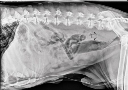
Figure 1: A lateral abdominal radiograph obtained after urinary catheter placement. Sublumbar lymphadenopathy is present (arrow), causing ventral displacement of the colon and compression of the rectum at the level of the pelvic canal.
Thoracic and abdominal radiography revealed mediastinal, intra-abdominal, and sublumbar lymphadenopathy, the latter causing ventral displacement of the colon and compression of the rectum at the level of the pelvic canal (Figure 1). Despite this finding, the patient did not demonstrate any difficulty defecating.
Because the patient was resistant to dorsal recumbency for abdominal ultrasonography despite sedation, general anesthesia was required. Ultrasonographic findings included severe lymphadenopathy of the sublumbar lymph nodes, irregular and markedly thickened bladder and urethral walls, and hypoechoic fat surrounding the neck of the urinary bladder and in the perirectal region (Figure 2). Additional findings included severe lymphadenopathy of the mesenteric lymph nodes, bilaterally hyperechoic renal cortices with reduced corticomedullary definition, two hypoechoic splenic nodules (measuring 18 mm and 13 mm), and a diffusely hypoechoic liver with course echotexture.

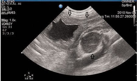

Figure 2. An ultrasonographic image showing enlarged sublumbar lymph nodes and irregular and markedly thickened bladder and urethral walls.
The ultrasonographic findings were suggestive of disseminated neoplasia. The differential diagnosis for the urethral obstruction in this patient was compression of the urethra from either sublumbar or subsacral lymphadenopathy or through direct neoplastic infiltration of the urethra, leading to the narrowing and occlusion of the lumen.
RECOMMENDATIONS TO OWNER
Recommendations to the patient's owner included fine-needle aspiration of abnormal abdominal organs and lymph nodes as well as a urine culture. The placement of a permanent urethral stent to help with urination was also offered. However, at the owner's request, the patient was euthanized.
POSTMORTEM CYTOLOGIC EXAMINATION
After euthanasia, ultrasound-guided fine-needle aspiration of the thickened bladder wall and the sublumbar lymph nodes was performed for cytologic evaluation. A population of monomorphic lymphoblasts was the predominant cell type in both samples, consistent with lymphoma. Additional testing (flow cytometry, polymerase chain reaction for antigen receptor rearrangement [PARR], immunohistochemistry) to distinguish B or T cell lymphoma was not performed.
DISCUSSION
Lymphoma is the most common hematopoietic neoplasm in dogs and generally presents in multicentric, gastrointestinal, mediastinal, and cutaneous forms. Less commonly, primary extranodal forms of the disease have been reported, including ocular, nasal, central nervous system, and cardiac.1 Lymphoma affecting the urinary bladder of dogs is rare, and only a few cases have been reported.2-6 More commonly, they are part of a multisystemic disease process, but primary extranodal urinary bladder lymphoma has also been documented. In two case reports of dogs with primary bladder lymphoma, one was B cell in origin and the other was T cell in origin.3,4
Urethral obstruction secondary to lymphoma affecting the lower urinary tract has been previously reported in two dogs, one with urethral lymphoma and the other with prostatic lymphoma. In the dog with urethral lymphoma, a well-marginated, midurethral tubular mass was the cause of the urinary obstruction. The patient also had a small intestinal mass, and T cell lymphoma was diagnosed on postmortem examination.7 The cause of the urinary obstruction in the dog with prostatic lymphoma was presumed to be infiltrative disease of the prostate, causing compression of the prostatic urethra.
Suction biopsy of the dog's urethra did not reveal neoplastic cells.8 Since a postmortem examination was not performed, it is difficult to determine whether this patient's urethral obstruction was due to neoplastic cells infiltrating the urethra causing intraluminal narrowing or was secondary to urethral compression by enlarged regional lymph nodes or the surrounding inflamed or neoplastic tissues.
In people
Lymphoma of the urinary bladder in people is a rare pathologic event that can either be a manifestation of widespread metastatic disease or solely affect the urinary bladder. In a retrospective study of patients with systemic non-Hodgkin's lymphoma, 10% to 20% had secondary involvement of the bladder at autopsy.9
Regarding primary bladder lymphomas, there are two predominant subtypes that have been equally documented: mucosa-associated lymphoid tissue (MALT) and a diffuse large B cell type. The MALT lymphoma subtype is thought to represent an unusual immune response to antigenic stimulation, as it appears to be associated with chronic cystitis from bacterial urinary tract infections.10,11 Histologically, the tumors are mostly considered non-Hodgkin's lymphoma of B cell type, and patients with these tumors often have an excellent prognosis as the tumors are considered low grade and are localized solely to the urinary bladder.
Patients with high-grade primary urinary bladder lymphomas of large B cell type are considered at diagnosis to have systemic disease.12 It has also been suggested that MALT lymphomas may transform into primary high-grade B cell lymphomas, but the proportion of tumors that undergo this transformation is not known.10,13 Although primary urinary bladder lymphomas are mostly B cell in origin, two cases of primary T cell lymphoma have been reported, one of which was suspected to occur secondary to chronic infection with shistosomiasis.14,15
Ultrasonography
The ultrasonographic appearance of canine urinary bladder lymphoma can be similar to findings reported with other bladder neoplasms and, thus, cannot solely be used to differentiate between them. Its appearance has been documented either as a heterogeneous mural mass that can affect all sites of the urinary bladder wall and potentially invade the serosa or as focal thickening of the urinary bladder wall.3,5 In addition to the regional changes within the urinary bladder, hydronephrosis and hydroureter have also commonly been reported.3,5
In people with urinary bladder lymphoma, the tumors most frequently present as a sessile solitary mass (66%) but can present as multiple sessile masses (14%), a polypoid mass (10%), or as diffuse thickening of the bladder wall (10%).14 In this case, the ultrasonographic appearance of the dog's bladder was consistent with a diffusely thickened and irregular wall and was not mass-like in appearance.
Clinical signs
Clinical signs that have been reported in dogs with lymphoma of the bladder include hematuria, stranguria, pollakiuria, dysuria, polydipsia, vomiting, diarrhea, weight loss, anorexia, and lethargy, in addition to no clinical signs at all.3-5 Intermittent hematuria, pollakiuria, and dysuria are the most frequent symptoms in people.10
Treatment options
Treatment of dogs with urinary bladder lymphoma may depend on whether the patient has multicentric lymphoma that has spread to the bladder or has primary malignant lymphoma that is limited to the urinary bladder. For patients with systemic disease, multiagent chemotherapy appears to be a logical treatment modality.
As primary urinary bladder lymphoma is relatively rare, there is little documentation of preferred modalities for treating it. In one case, hypofractionated external beam radiation and cytotoxic chemotherapy was used to treat a 3-year-old spayed mixed-breed dog with a large infiltrative lymphomatous mass encompassing two-thirds of the bladder lumen. The patient achieved complete remission and had been in remission for 52 months when that case report was published.3
Limitations with this case
A limitation of this case report is that the patient was euthanized before reinstituting therapy. Also, the owner did not consent to a necropsy examination to assess the full extent of disease. It is unfortunate that neither histology nor specific testing to distinguish B cell from T cell lymphoma was performed either before or after referral. Had the client opted to pursue additional treatment, restaging the patient, placing a urethral stent, and starting a rescue chemotherapy protocol in addition to radiation therapy would have been considered.
CONCLUSION
This report documents an unusual presentation of urinary bladder lymphoma in a dog with a urinary obstruction. While cases of urinary bladder lymphoma have been previously described in the literature, this case is the first report of confirmed multicentric lymphoma with bladder involvement being associated with a urinary obstruction. Urinary bladder lymphoma should be included as a differential diagnosis for urinary obstructions in dogs, especially in dogs with multicentric lymphoma.
REFERENCES
1. Vail D, Pinkerton M, Young K. Canine lymphoma and lymphoid leukemias. In: Withrow S, MacEwen E, eds. Small animal clinical oncology. 5th ed. Philadelphia, Pa: W.B. Saunders, 2012;608-637.
2. Strafuss AC, Dean MJ. Neoplasms of the canine urinary bladder. J Am Vet Med Assoc 1975;166:1161-1163.
3. Kessler M, Kandel-Tschiederer B, Pfleghaar S, et al. Primary malignant lymphoma of the urinary bladder in a dog: longterm remission following treatment with radiation and chemotherapy. Schweiz Arch Tierheilkd 2008;150:565-569.
4. Maiolino P, DeVico G. Primary epitheliotropic T-cell lymphoma of the urinary bladder in a dog. Vet Pathol 2000;37:184-186.
5. Benigni L, Lamb CR, Corzo-Menendez N, et al. Lymphoma affecting the urinary bladder in three dogs and a cat. Vet Radiol Ultrasound 2006;47:592-596.
6. Van Noort R. [4 unusual cases of malignant lymphoma in the dog and cat]. Tijdschr Diergeneeskd 1997;122:502-505.
7. Struble AL, Lawson GW, Ling GV. Urethral obstruction in a dog: an unusual presentation of T-cell lymphoma. J Am Anim Hosp Assoc 1997;33:423-426.
8. Winter MD, Locke JE, Penninck DG. Imaging diagnosis-urinary obstruction secondary to prostatic lymphoma in a young dog. Vet Radiol Ultrasound 2006;47:597-601.
9. Sufrin G, Keogh B, Moore H, et al. Secondary involvement of the bladder in malignant lymphoma. J Urol 1977;118,251-253.
10. Bates A, Norton A, Baithun S. Malignant lymphoma of the urinary bladder: a clinicopathological study of 11 cases. J Clin Pathol 2000;53:458-461.
11. Ohsawa M, Aozasa K, Horiuchi K, et al. Malignant lymphoma of bladder: report of three cases and review of the literature. Cancer 1993;72:1969-1974.
12. Coskun U, Günel N, Eroglu A, et al. Primary high grade malignant lymphoma of bladder. Urol Oncol 2002;7:181-183.
13. Isaacson P. Critical commentary to primary malignant lymphoma of the bladder. Path Res Pract 1996;192:164-165.
14. Choi JH, Jeong YY, Shin SS, et al. Primary calcified T-cell lymphoma of the urinary bladder: a case report. Korean J Radiol 2003;4:252-254.
15. Mourad WA, Khalil S, Radwi A, et al. Primary T-cell lymphoma of the urinary bladder. Am J Surg Pathol 1998;22:373-377.