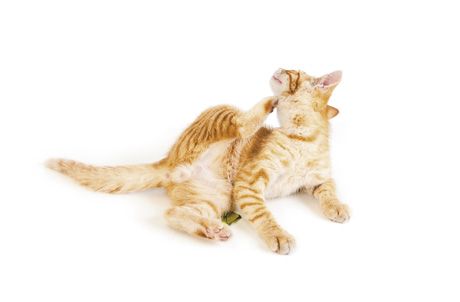
CVC Highlight: Getting to the bottom of feline facial dermatitis
What to do when your veterinary client exclaims, My cat has something wrong with his face!
Feline facial dermatoses represent a reasonably common clinical complaint but a wide variety of underlying diseases. Here is an overview of the many disease syndromes that can cause facial dermatitis in cats.
Parasites
Facial dermatitis related to a parasite is most commonly caused by a mite infestation rather than something more common such as fleas or cheyletiellosis. Feline scabies (Notoedres cati) is rare but causes an extremely pruritic dermatitis of the face and neck. One of the newly described Demodex species mites, such as gatoi or the “unnamed” mite, may cause contagious, pruritic facial dermatitis. These mites are easily found and mostly easily treated.
In the southern United States, cutaneous hypersensitivity reactions to mosquito bites are occasionally seen-typically as ulcerative to plaque-like lesions on the hairless portion of the nose. Although difficult to diagnose at first, these lesions will abate with brief corticosteroid treatment and restriction to indoors.

Infections
Infections are not a common primary cause of dermatitis restricted to the face, with the strong exception of dermatophytosis. Nevertheless, a cytologic examination should be performed to check for any bacterial or yeast overgrowth, which is typically secondary. A Wood's lamp examination and fungal culture are mandatory in all cases of facial dermatitis, no matter the appearance.
The other important infectious cause of facial dermatitis is feline herpesvirus 1 (FHV1). Although rare, this aberrant infection causes severe ulcerative, necrotic erosions to plaque-like lesions on the face that may be mistaken for other diseases such as eosinophilic granuloma. Generally the virus can be found by direct histologic examination, immunohistology, or PCR testing of the biopsy tissue. This is a good example of why it is important for any cat with persistent, rather severe, ulcerative facial dermatitis to undergo a biopsy. In addition to providing a specific diagnosis, finding FHV1 allows you to provide specific and effective treatment; famciclovir at 125 mg/cat orally b.i.d. for at least 21 days has proved very effective.
Allergic disease
In theory, food, environmental, and insect allergies can all manifest as facial dermatitis. Of these, a food allergy should be a primary suspect in feline head and neck pruritus. Dietary restriction and provocation trials are mandatory to make a diagnosis.
Pemphigus foliaceus
As the most common autoimmune skin disease in cats, pemphigus foliaceus typically has a strong component of facial distribution, particularly on the bridge of the nose, around the eyes, and on the ear pinnae. Most cats also have involvement of their footpads or nailbeds (caseous paronychia), but this is not always the case. Many cats with pemphigus foliaceus will have a prolonged history of cycles of improvement and worsening clinical signs. The clinical appearance of pemphigus foliaceus is rather dramatic and unusual, but it is important to confirm this disease by performing a biopsy.
There have been reports of pemphigus foliaceus-like disease in cats caused by primary infections, particularly with dermatophytes. Because of the waxing and waning nature of pemphigus foliaceus, you may mistake your empirical treatment for a different disease as successful.
Feline acne
There is much myth and legend about the cause of feline acne. Most cases are probably idiopathic, perhaps with a cause similar to that in people-defective cornification in the follicle or a sebaceous duct leading to plugging with keratinaceous debris and secondary infection. Rarely, a defined cause such as primary bacterial infection or dermatophytosis may be responsible. Causes such as the type of food bowl, food allergy, and failure of chin grooming are much more speculative, and there is no convincing evidence for this pathogenesis.
Treatment relies initially on clearing any secondary bacterial infection with antibiotics and then providing daily facial hygiene with keratolytic topical products (2% to 3% benzoyl peroxide or salicylic acid), which may need to be done periodically on a long-term maintenance basis. For recalcitrant cases, I have had success with topical tretinoin gel or cream (0.025% applied twice daily until resolution and then once every one to two days to maintain remission if needed).
“Dirty face” in Persian cats is an uncommon but frustrating idiopathic facial dermatitis that is considered by some to be a more severe form of feline acne. In this disease, comedones and crusts extend beyond the chin area to the facial folds and preauricular areas. Diagnostic evaluations should be performed to rule out definable causes, but most cases are idiopathic, and a genetic alteration may exist in this breed. Therapy is symptomatic, using topical products as for feline acne.
Indolent ulcers
Indolent (“rodent”) ulcers appear as well-demarcated ulcers with raised borders present on the margin of the upper or lower lip. Initial diagnosis is generally straightforward, based on clinical appearance and cytology: remove surface debris with a gauze pad to expose the moist underlying tissue, and then take impression smears. Finding eosinophils on cytologic examination is sufficient to make an initial tentative diagnosis. Note that if cocci are also found, especially within the inflammatory cells, first treat the cat with antibiotics since this disease can sometimes reflect an aberrant response to bacterial infection.
In an otherwise healthy young animal, it is acceptable to treat a cat with one course of injectable corticosteroids (20 mg methylprednisolone acetate every two weeks for a total of three injections). If the disease is especially severe, recalcitrant, or recurrent, search for an underlying cause (including biopsy with assessment for FHV1 presence), and avoid repeated corticosteroid use. For recalcitrant cases, cyclosporine at 7 to 10 mg/kg once daily until remission, then tapering to a minimum maintenance dose, is often effective.