Dermatology Challenge: Resolution of a necrotizing wound in a debilitated cat
A 6-year-old, 11.2-lb (5.1-kg), neutered male domestic shorthaired cats was presented to South Athens Animal Clinic for evaluation of a large cutaneous wound.
A 6-year-old, 11.2-lb (5.1-kg), neutered male domestic shorthaired cat was presented to South Athens Animal Clinic for evaluation of a large cutaneous wound. The cat had been absent from its residence for nine days. Before its disappearance, the cat had been in good health, and its vaccination status, including rabies, was current.
Physical examination and diagnostic procedures
At presentation, the cat was listless with a subnormal body temperature of 100 F (37.7 C). The cat was determined to be about 10% dehydrated based on prolonged skin tenting and delayed capillary refill time. A 3-x-4-in cutaneous wound in the left axilla extended over most of the scapula, including the lateral and ventral thorax and upper forelimb.
Initial evaluation revealed a necrotizing, subcutaneous lesion involving the fascia and superficial musculature of the lateral shoulder, thorax, and triceps area. The thoracic cavity and deeper musculature exhibited no penetrating wounds or tears, but portions of the dorsal and lateral tendons attaching muscle groups to the olecranon were exposed. The elbow joint capsule and tendon sheaths of the triceps, anconeus, and ulnaris lateralis muscles were visible but not compromised. The skin on the medial side of the left forelimb was intact. In addition to the primary wound site, two superficial puncture wounds were detected on the cat's right lumbar area and thorax, leading to a presumptive diagnosis of a chronic, infected bite wound. Results of feline leukemia virus and feline immunodeficiency virus ELISA tests were negative. Additional blood work was not done because of the cat's debilitated state. A decision about continuing treatment would be made after wound débridement.
Treatment
After the physical examination, the cat was anesthetized by intravenous administration of tiletamine hydrochloride and zolazepam hydrochloride (3 mg/kg) in combination with 0.06 mg/kg acepromazine maleate. Débridement consisted of first gently flushing the wound with lactated Ringer's solution. The skin over the left chest wall, forelimb, axilla, and shoulder was also shaved. Obviously devitalized tissue was surgically excised. Because of the wound size, we were conservative with tissue removal.
The resulting open wound was flushed several times with lactated Ringer's solution, and wet-to-dry bandages were applied to the left side of the thorax and left forelimb. The area surrounding the two superficial puncture wounds on the right side was shaved, and the wounds were treated with a topical antibiotic ointment. Lactated Ringer's solution was administered slowly intravenously (240 ml/day), and liquefied food (Prescription Diet a/d—Hill's, mixed with water) was given orally by syringe. Amoxicillin (10 mg/kg orally b.i.d.) was initiated.
The cat was anesthetized again the next day to remove residual necrotic tissue, and the wound was débrided a second time, resulting in a larger area (8 x 8 in) of exposed subcutaneous fascia and superficial musculature (Figure 1). The area was copiously flushed with lactated Ringer's solution, and wet-to-dry bandages were applied. The cat's prognosis was guarded because of the extent of the wound and the cat's debilitated condition.

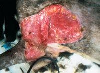

Figure 1: The appearance of the wound after the second débridement (Day 2).
On Day 3, 1 L of an aqueous solution of EDTA-tris (8 mmol USP disodium ethylenediamine-tetraacetate dihydrate and 20 mmol USP 2-amino-2-hydroxymethyl-1,3-propanediol; Tricide—Molecular Therapeutics, Athens, Ga.) was mixed with 125 mg ampicillin; the mixture was used to flush and cleanse the tissues. Gauze bandages, soaked in the Tricide–ampicillin solution, were placed over the wound. To prevent the bandages from drying out and sticking to the wound, the gauze covering the chest wall and left forelimb was covered with food-grade plastic wrap, which was held in place with loosely applied Vetrap (3M). Additional gauze and light bandaging were applied to hold the wet bandages and plastic wrap in place.
On Day 4, the cat's body temperature had increased to 104 F (40 C), and we noted peripheral edema in the left forepaw. The bandages were repositioned to relieve the pressure on the distal forelimb. The cat remained depressed but was able to sit up and move around in its cage and was eating small quantities of liquefied food. Because of this increased activity and the frequent bandage changes, hydration was maintained by subcutaneous administration of lactated Ringer's solution (120 ml b.i.d.). We performed a radiographic examination of the thorax (to rule out pneumonia) and abdomen (to rule out fecal impaction). The thoracic radiographs were normal. The abdominal radiographs revealed a diffuse, intraluminal soft tissue opacity causing colonic distention. The cat received two soap-and-water enemas that day, which resulted in the passage of a large quantity of feces. The cat was also given lactulose syrup (0.67 g [1 ml] orally b.i.d. for 21 days), orbifloxacin (4.5 mg/kg orally q.i.d. for 10 days), metronidazole (22 mg/kg orally b.i.d. for seven days), and butorphanol tartrate (0.2 mg/kg orally b.i.d.).
The cat remained hospitalized for the next five days and was given liquefied food orally and lactated Ringer's solution subcutaneously (120 ml b.i.d.). Wound treatment consisted of flushing the site daily with 100 to 200 ml Tricide-ampicillin solution and applying wet bandages covered with plastic wrap and Vetrap as described above. Cage rest and supportive care continued, and the cat's body temperature returned to normal by Day 9.


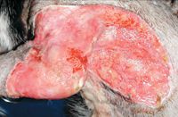
Figure 2: Marked granulation is present over most of the exposed tissue (Day 8).
On Day 8, the wound had sufficient granulation tissue to support surgical closure of the thoracic and scapular regions of the wound (Figure 2), which was performed the next day. Anesthesia was induced with tiletamine and zolazepam and maintained with 2% isoflurane. To close the wound, the skin margins were freshened, and the skin on the ventral thorax and lateral shoulder was extensively undermined. These two sections were pulled together over the olecranon. The dorsal and ventral margins were apposed and sutured with 3-0 polydioxanone in a simple interrupted pattern (Figure 3). A Penrose drain was placed along the left lateral thoracic wall, which exited at the ventral margin of the surgical site. The cat recovered from anesthesia without complications.


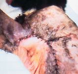
Figure 3: Surgical closure of the thoracic and scapular area (Day 9). Suture tension required subsequent removal of four sutures, which partially opened the wound to allow forelimb movement. The arrows indicate the site of the drain exit.
Postoperatively, excessive skin tension at the suture line over the elbow was noted. This finding required that the cat be anesthetized again later that day. The suture line was opened by removing four sutures from the point of the elbow dorsally to the intersection of the shoulder and lateral chest wall. This left about 10% of the surgical site open and exposed portions of the tendons, fascia, and granulation bed over the elbow. Postoperatively, hot packs were placed on the surgical site two or three times a day to enhance skin attachment to the granulation bed as well as to improve local circulation. The exposed tissues were rinsed with the Tricide–ampicillin solution, and the wound was left open to heal by second intention.
The cat was quite active, so the drain was lightly covered with gauze and one layer of Vetrap. The drain fluid remained clear, and there was little exudate (1 to 2 ml per bandage change). On Day 11, a complete blood count and serum chemistry profile were done because the cat was not gaining weight; it weighed 10.6 lb (4.8 kg). The only abnormalities were a moderate neutrophilia (27.7 × 10³ neutrophils/μl; normal = 2.5 to 12.5 × 10³/μl) and a mild hypoproteinemia (total protein 5.4 g/dl; normal = 6 to 7.77 g/dl) with hypoalbuminemia (albumin 2 g/dl; normal = 3 to 4.3 g/dl). These results indicated inflammation and protein loss from the wound without sufficient protein replacement by oral feeding. Although the cat did not object to syringe feeding, it was not consuming enough food to maintain its weight, so it was discharged and allowed to recover at home. The cat was given a third enema before discharge.
Home care and follow-up
Daily in-home supportive care consisted of subcutaneously administering lactated Ringer's solution (120 ml b.i.d.), administering the prescribed oral medications (amoxicillin and lactulose), applying hot packs, and washing the suture lines with the Tricide-ampicillin solution. Fluids were administered for one additional week while antibiotic therapy continued for two weeks. To prevent the exposed subcutaneous tissues of the elbow and proximal limb from drying out, about 5 ml of an emulsion containing Tricide, liquid lanolin, vitamin E oil, ampicillin, and deionized water was applied to the affected area twice a day. The emulsion was prepared by mixing 35 ml lanolin and 5 ml vitamin E (333 IU/ml) with 10 ml deionized water that contained 60 mg of Tricide (1.2 mg/ml) and 0.5 mg/ml ampicillin. Light bandaging of the shoulder and forelimb restricted movement, irritated the suture line, and was eventually removed by the cat.
On Day 15, the cat was returned to the hospital for reevaluation. The suture lines were intact and the skin over the thorax and scapular regions had attached to the underlying granulation bed. The skin on the ventral thorax and axilla, however, had not reattached because of forelimb movement. About half of the exposed elbow and upper limb tissue had formed a granulation bed, and the wound margins appeared healthy. We removed the drain and decided to continue at-home wound management, including administering the amoxicillin, lactulose, and topical Tricide emulsion and placing hot packs.

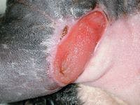

Figure 4: The contracted wound before closure six weeks after initial presentation (Day 40).
The cat was readmitted to the hospital for examination 25 days later (Day 40). After three and a half weeks of home care, the open wound over the elbow had constricted to about 1.5 x 2 in (Figure 4), and the skin covering the axilla had reattached except for a subcutaneous pocket on the lateral thoracic wall. This small pocket was about 1.5 x 2.5 in and was medial to the olecranon. On Day 44, the cat was anesthetized to close the open wound. The margins of the wound were freshened, the edges were minimally undermined, and the area was sutured in a simple interrupted pattern. The cat was discharged the next day, and the sutures were removed nine weeks after the initial presentation (Day 63) (Figure 5).


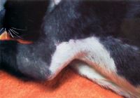
Figure 5: The affected area after suture removal (Day 63).
Discussion
Bite wounds are common in outdoor cats. Short-term lesions often respond favorably to minimal wound care and systemic antibiotics, but contaminated wounds such as the one described in this case report may develop secondary complications, such as necrotizing dermatitis, fasciitis, or myositis. Aerobic and anaerobic bacteria within devitalized tissue may interact synergistically to induce deep tissue cellulitis or abscessation, resulting in draining sinuses or skin sloughing.1 This report details the successful management of a chronic wound in a debilitated cat in which limb amputation, death, or euthanasia were possible sequelae. Because of the severity and chronic nature of the wound, postoperative complications were expected.
Formulating a prognosis for large, necrotizing wounds in dogs or cats requires considering multiple factors, including the animal's physical condition, the severity and extent of the wound, and the client's commitment to pursuing treatment. Any treatment that facilitates regeneration of healthy tissue and improves wound healing may turn a patient presented for euthanasia into one that has a reasonable chance of recovery. Initiating systemic antibiotic treatment and supportive care and properly treating the open wound during the first few days of hospitalization allowed sufficient time for this cat to stabilize and begin showing signs of recovery.


A fully recovered Oscar, the cat featured in this case report, in 2004.
After débridement, a primary issue was how to control infection and prepare a large area of exposed subcutaneous tissue for surgical closure. In this contaminated bite wound, we chose delayed closure because there was concern regarding tissue viability and residual areas of infection.2 We added Tricide supplemented with ampicillin to the treatment regimen to facilitate pathogen control and wound health, which allowed surgical closure eight days after presentation. Granulation tissue usually appears three to six days after injury in nondebilitated patients.3
While the wound was open, the only treatment capable of protecting such a large area of exposed tissue was wet bandaging. While the cat was hospitalized, the wound was flushed with the Tricide-ampicillin solution, and wet bandages soaked in the same solution were applied daily. Frequent bandage changes allowed the wound to be examined for signs of healing or development of additional necrotic areas. Wet bandaging protects exposed tissues while maintaining cellular hydration. A potential disadvantage of wet bandaging is nosocomial wound contamination.4 Applying bandages soaked in low concentrations of chlorhexidine (0.05%) can reduce the number of bacteria entering an open wound but may not control heavy Pseudomonas species infections.5 Higher concentrations of chlorhexidine may kill bacteria more effectively but also tend to delay the formation of granulation tissue.5
Previous in vitro testing has shown that the active ingredients in Tricide have an antibacterial effect when applied directly to wounds or bandages.6,7 These compounds in combination with various antibiotics have also been shown to reduce the minimum inhibitory concentrations of antibiotics capable of killing bacteria found in contaminated wounds (i.e.Pseudomonas species, Staphylococcus species, and Escherichia coli).8,9 The mechanism of action is thought to be increased uptake of antibiotics by the bacteria, thus enhancing the bactericidal effect.10 In this case, systemic antibiotics and repeated flushing of the open wound with Tricide resulted in excellent wound healing. A healthy granulation bed was evident within a week of initiating treatment, thereby allowing surgical closure. After surgery, topical treatments of the area included applying a mixture of lanolin, vitamin E, Tricide, and ampicillin. The decision to use Tricide in this case was based on in vitro and in vivo documentation of its ability to enhance antimicrobial agent activity.11-18
After surgical closure and opening the suture line over the elbow, wet bandaging of the proximal forelimb and olecranon was not possible because of limb movement. Since the cat was being discharged to its home environment, maintaining tissue hydration and preventing infection were important. Incorporating the Tricide–ampicillin mixture into a lanolin and vitamin E emulsion proved to be a satisfactory course of treatment. After three and a half weeks of topical treatment, the wound had contracted considerably, and a healthy granulation bed had formed over most of the wound's surface. The surrounding skin and subcutaneous tissue remained pliable, thus contributing to successful surgical closure and clinical resolution.
On presentation, the clinical assessment of this case was guarded with a poor prognosis for complete recovery. Although the degree of superficial tissue damage was extensive, the lack of penetrating wounds or fractures contributed to this patient's chances for improvement. Exposure of the tendons of the forelimb and surrounding tissue delayed closure and increased the risk of infection. We chose healing by second intention since wound closure over points of high skin tension, such as in this case, cannot be relied on because of poor underlying cutaneous blood supply.19 With wound contraction, surgical closure proved feasible three and a half weeks later.
Clinical resolution of this challenging case was possible because of a lack of complications during wound healing and postsurgical closure. Under such conditions, motivated owners capable of treating their cats at home may elect similar treatments and thereby avoid euthanasia or shorten the hospitalization of their pets.
ACKNOWLEDGMENTS
The authors would like to thank the technicians and staff of South Athens Animal Clinic for their excellent care of Oscar. They would also like to acknowledge the valuable input of Branson W. Ritchie, DVM, PhD; and Richard E. Wooley, DVM, PhD, of the University of Georgia College of Veterinary Medicine for technical advice in the use of Tricide and wound management.
The photographs and information for this case were provided by Joanne Maki, DVM, PhD, Department of Medical Microbiology, College of Veterinary Medicine, University of Georgia, Athens, GA 30602; and Mark Mosher, DVM; and Thomas Nemetz, DVM, PhD, South Athens Animal Clinic, 2040 S. Milledge Ave., Athens, GA 30606. Dr. Maki's current address is 3221 Smithonia Road, Colbert, GA 30628.
REFERENCES
1. Swaim, S.F.: Specific types of wounds—Bite wounds. Small Animal Wound Management (S.F. Swaim; R.A. Henderson, eds.). Williams & Wilkins, Baltimore, Md., 1997; pp 112-117.
2. Holt, D.E.; Griffin, G.: Bite wounds in dogs and cats. Vet. Clin. North Am. (Small Anim. Pract.) 30 (3):669-679; 2000.
3. Swaim, S.F.: Wound healing. Small Animal Wound Management (S.F. Swaim; R.A. Henderson, eds.). Williams & Wilkins, Baltimore, Md., 1997; pp 1-12.
4. McManus, W.F. et al.: Burn wound infection. J. Trauma 21 (9):753-756; 1981.
5. Swaim, S.F.: Wound dressing materials and topical medications. Small Animal Wound Management (S.F. Swaim; R.A. Henderson, eds.). Williams & Wilkins, Baltimore, Md., 1997; pp 53-85.
6. Roberts, N.A. et al.: The bactericidal action of ethylenediaminetetra-acetic acid on Pseudomonas aeruginosa. Microbios 2 (7-8):189-208; 1970.
7. Wooley, R.E.; Jones, M.S.: Action of EDTA-Tris and antimicrobial agent combinations on selected pathogenic bacteria. Vet. Microbiol. 8 (3):271-280; 1983.
8. Wooley, R.E. et al.: In vitro action of combinations of antimicrobial agents and EDTA-tromethamine on Escherichia coli. AJVR 44 (6):1154-1158; 1983.
9. Wooley, R.E. et al.: In vitro action of combinations of antimicrobial agents with EDTA-tromethamine on Proteus vulgaris of canine origin. AJVR 45 (7):1451-1454; 1984.
10. Wooley, R.E. et al.: Uptake of antibiotics in gram-negative bacteria exposed to EDTA-Tris. Vet. Microbiol. 10 (1):57-70; 1984.
11. Goldschmidt, M.C. et al.: EDTA and lysozyme lavage in the treatment of pseudomonas and coliform bladder infections. J. Urol. 107 (6):969-972; 1972.
12. Blue, J.L. et al.: Treatment of experimentally induced Pseudomonas aeruginosa otitis externa in the dog by lavage with EDTA-tromethamine-lysozyme. AJVR 35 (9):1221-1223; 1974.
13. Youngquist, R.S.: Pseudomonas metritis in a mare. VM/SAC70 (3):340-342; 1975.
14. Wooley, R.E. et al.: Antibiotic-tromethamine-EDTA lavage for the treatment of bacterial rhinitis in a dog. JAVMA 175 (8):817-818; 1979.
15. Bjorling, D.E.; Wooley, R.E.: EDTA-tromethamine lavage as an adjunct treatment for multiple fistulas in a dog. JAVMA 181 (6):596-597; 1982.
16. Youngquist, R.E. et al.: The effects of EDTA-Tris infusion on the equine endometrium. Theriogenology 22:593-599; 1984.
17. Sparks, T.A. et al.: Antimicrobial effect of combinations of EDTA-Tris and amikacin or neomycin on the microorganisms associated with otitis externa in dogs. Vet. Res. Commun. 18 (4):241-249; 1994.
18. Wooley, R.E. et al.: In vitro evaluation of the antimicrobial effect of commercially available mastitis medications combined with EDTA-Tris on bacteria that cause mastitis in cattle. Vet. Ther. 3:1-7; 2002.
19. Probst, C.W.: Grafting techniques and failures in small animals. Problems in Veterinary Medicine: Reconstructive Surgery (W.A. Lindsay, ed.). J.B. Lippincott, Philadelphia, Pa., 1990; pp 413-423.