Exploring your digital radiography equipment options
One of the first steps in switching to digital radiography is deciding which type of system to purchase.
One of the first steps in switching to digital radiography is deciding which type of system to purchase. Two broad classes of digital radiography equipment are available for veterinarians: computed radiography (CR), which is cassette-based, and direct (capture) radiography (DR).1,2
This article focuses on primary capture devices instead of secondary capture devices such as image digitizers (scanners) and digital cameras. In general, secondary capture devices have important limitations from both a medicolegal and an image quality standpoint.

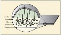

1. A schematic of traditional film in a double-screen cassette system. X-ray photons pass through the cassette to interact with screens housed on both sides of the film. When an X-ray photon interacts with the screen, light is emitted instantaneously, which then exposes the film. (Illustrations by John H. Doval.)
CR
CR was the first digital radiography system used in medical imaging. CR systems have many features analogous to those of traditional film-screen systems (Figure 1) because a cassette is used to house a photo-stimulable phosphor sheet.2 CR radiographs are obtained in a traditional manner: all the views of the study are taken, and then the cassettes are moved en masse to a digital processor. Each cassette is then individually fed into the processor where the storage phosphor sheet is removed and scanned by a laser (Figure 2). The laser imparts energy onto the storage phosphor sheet, liberating light from the latent image stored therein. The light emitted from the phosphor sheet is digitized to produce the digital radiograph.
The processor requires 60 to 90 seconds to produce an image from each cassette. During this processing time, the next cassette cannot be fed into the processor, though many processors accept multiple cassettes (two to four) at one time. CR may even slow workflow if the number of radiographic projections exceeds the number of cassette receptacles on the processor.3 This problem may arise in multiple-view studies such as orthopedic or pre-purchase examinations.

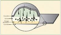

2. A schematic of a typical CR cassette. X-ray photons pass through the cassette to interact with the storage phosphor. The X-ray energy is stored in the phosphor sheet. When the cassette is inserted into the processor, a laser passes over the storage phosphor, releasing the stored energy as light. The computer processes this light energy to yield a digital image.
Thus, the CR workflow is similar to that of traditional radiographic film.4,5 Because of the delay in image processing, it will take several minutes to identify errors in radiographic techniques, specifically positioning. For small-animal practice, this may simply entail repositioning the animal. But mobile practitioners won't recognize the need for retakes until the cassettes are returned to the processor, which is most commonly housed in a stationary location such as the clinic.
CR requires purchasing the cassettes, a processor, and equipment for viewing and storing the images (see the article "Getting the most out of digital image viewing" and the article "How to store digital images and comply with medical recordkeeping standards" ). The cassettes have the same physical dimensions as film-based cassettes and do not require any modification to the X-ray generator, tables, or grids. In general, CR is a slightly less expensive digital radiographic modality that will provide many of the advantages of digital imaging in a veterinary practice.
DR
DR, or direct X-ray (DX), generally refers to one of three system types. The unifying characteristics of these systems are that an image is produced nearly instantaneously and that the same X-ray detector is used for every exposure.1,2,6-8 Two DR systems fall into the flat panel category. Flat panel detectors are a commonly used DR system in veterinary medicine. The other type of DR system is a charge-coupled device (CCD) camera.
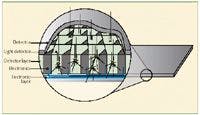
3. A schematic of a scintillation style, flat panel DR detector. X-ray photons pass through the detector and interact with a scintillation crystal that emits light. Each pixel of the detector houses electrical hardware, which maps the light as it is emitted.
Flat panel detectors
The DR flat panel detectors are further subdivided into panels that scintillate (i.e. emit light energy that is digitized and recorded when X-ray photons interact with the detector elements) (Figure 3) and panels that create an electrical current when X-ray photons interact with the detector; the current is then converted to a radiographic image.9 In either case, this process is nearly instantaneous, and images can generally be viewed within seconds of their acquisition. Scintillation detectors are currently the most prevalent veterinary DR system.10 Theoretically, in systems that do not scintillate, the lack of the intermediate light-producing step should provide these systems with superior resolution. The clinical significance of this difference has yet to be determined.1,7-9,11
The various vendors' panels have different physical dimensions, but all have a slightly smaller active detection area than the overall dimensions of the panel. The size of the radiograph is limited to the detector size, and care should be taken to choose the correct imaging surface size for the types of patients and studies being produced. Generally, the dimensions of this type of detector will be different from conventional film-screen cassettes, occasionally requiring alterations in radiographic examination tables or preexisting cassette holders (equine practice). Furthermore, to electronically synchronize the DR system with the existing generator, a minor system modification, which is usually done by the vendor, is necessary. As with CR systems, both flat panel and CCD DR systems require purchasing a means of image storage and review.
CCD cameras
CCD camera radiographic systems generate an X-ray image when X-rays interact with a fluorescent (light-producing) screen that is attached to the underside of the X-ray table. The light produced from this interaction is then focused by a lens and captured by the CCD chip. In essence, the CCD camera photographs the light produced from X-rays interacting with a screen.
CCD chips are relatively small (2.5 x 2.5 cm to 8 x 8 cm). The large demagnification factor needed to reduce the size of the patient's image to the size of the CCD may result in loss of information, which has limited the quality of CCD-based images in the past.2,10 Advances in CCD technology, however, have overcome this limitation, and CCD-based machines are now accepted in human radiographic applications.12-15 The veterinary experience with CCD cameras is more limited than with CR and flat panel DR detectors. CCD radiography is used extensively in human and veterinary dental radiography because of the small field of view.
CONCLUSION
Both CR and DR systems create a set of raw data that will be used to produce a diagnostic radiographic image. Each vendor has a different method of image processing that may involve several steps that vary in complexity and involve mathematical manipulations of the raw data. Although there are marked technical differences among various vendors' radiographic systems, differences in image quality stem mainly from a system's ability to effectively process the image and not necessarily from the type of equipment used to obtain the image. CR is a relatively inexpensive system of digital radiography that shares a similar workflow with film-based radiography. DR technologies enable veterinarians to generate radiographic images within seconds of acquisition.
Sarah M. Puchalski, DVM, DACVR
Department of Surgical and Radiological Sciences
School of Veterinary Medicine
University of California
Davis, CA 95616
REFERENCES
1. Bruce R. CR versus DR—what are the options? AuntMinnie, 2003. Available at: www.auntminnie.com/index.asp?sec=ser&sub=def&pag=dis&ItemID=58851.
2. Bushberg JT, Seibert JA, Leidholdt Jr, EM, et al. Digital radiography. In: The essential physics of medical imaging. 2nd ed. Philadelphia, Pa: Lippincott, Williams & Wilkins, 2002.
3. Puchalski S, Hornof W. A limited image quality comparison of film-screen, computed radiography and digital radiography, in Proceedings. Am Coll Vet Radiogr Annu Mtg 2004.
4. Andriole KP. Productivity and cost assessment of computed radiography, digital radiography, and screen-film for outpatient chest examinations. J Digit Imaging 2002;15:161-169.
5. Andriole KP, Luth DM, Gould RG. Workflow assessment of digital versus computed radiography and screen-film in the outpatient environment. J Digit Imaging 2002;15(suppl 1): 124-126.
6. Digital x-ray systems. Part 1. An introduction to DX technologies and an evaluation of cassette DX systems. Health Devices 2001;30;273-310.
7. Digital x-ray systems. Part 2. An overview of digital radiography concepts and an evaluation of cassetteless DX systems. Health Devices 2001;30:381-401.
8. Fischbach F, Ricke J, Freund T, et al. Flat panel digital radiography compared with storage phosphor computed radiography: assessment of dose versus image quality in phantom studies. Invest Radiol 2002;37:609-614.
9. Goo JM, Im JG, Kim JH, et al. Digital chest radiography with a selenium-based flat-panel detector versus a storage phosphor system: comparison of soft-copy images. AJR Am J Roentgenol 2000;175:1013-1018.
10. Hornof W. Digital radiography: The available technologies and how to separate hype from reality, in Proceedings. Am Vet Med Assoc Annu Convention 2005.
11. Peer S, Neitzel U, Giacomuzzi SM, et al. Direct digital radiography versus storage phosphor radiography in the detection of wrist fractures. Clin Radiol 2002;57:258-262.
12. Kroft LJ, Geleijns J, Mertens BJ, et al. Digital slot-scan charge-coupled device radiography versus AMBER and Bucky screen-film radiography for detection of simulated nodules and interstitial disease in a chest phantom. Radiology 2004;231:156-163.
13. Kroft LJ, Veldkamp WJ, Mertens BJ, et al. Comparison of eight different digital chest radiography systems: variation in detection of simulated chest disease. AJR Am J Roentgenol 2005;185:339-346.
14. Veldkamp WJ, Kroft LJ, Boot MV, et al. Contrast-detail evaluation and dose assessment of eight digital chest radiography systems in clinical practice. Eur Radiol 2006;16:333-341.
15. Veldkamp WJ, Kroft LJ, Mertens BJ, et al. Digital slot-scan charge-coupled device radiography versus AMBER and Bucky screen-film radiography: comparison of image quality in a phantom study. Radiology 2005;235:857-866.