Fatal skin diseases in dogs and cats: What veterinary professionals need to know
They're rare, unusual and prognosis is often dismal. But in the event you encounter one of these difficult dermatologic cases, you'll want to be prepared. Heres the latest on potentially fatal skin diseases in small animals.


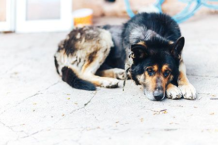
Anastassiya/stock.adobe.com
No, skin diseases in small animals aren't typically fatal.
However, the lesions associated with some skin diseases in dogs and cats are severe enough to make a pet look so bad (not to mention, feel) that its owner no longer wants to deal with the disease and decides to relinquish-or even euthanize-the pet.
Stephen D. White, DVM, DACVD, professor of medicine and epidemiology at the University of California-Davis, recently spoke about the skin conditions that threaten the well-being of the affected animal. Here are his insights on neoplastic, metabolic, immune-mediated and neutrophilic small animal skin diseases.
Neoplastic skin disease
Feline paraneoplastic alopecia, a ventral alopecia of older cats, is usually acute in onset, rapidly progressive and symmetrical, affecting the ventrum and limbs. Affected cats groom excessively and have characteristic glistening (but not fragile) abdominal skin. Footpads can also be affected with crusts, fissures and sloughing.1,2 A secondary Malassezia infection may be present. Histopathology reveals severe hair follicle atrophy and miniaturization.1,2 Diagnosis can be made with thoracic radiographs but is most commonly made with abdominal ultrasound.
This disease is associated with exocrine pancreatic adenocarcinoma. Unfortunately, once the alopecia is noticed, the cancer has likely already metastasized. Removing the primary tumor provides only temporary relief. In addition, removing the pancreas can lead to diabetes.
In short, feline paraneoplastic alopecia does not have a good prognosis.
Metastatic pulmonary carcinomas occur in cats and affect the distal extremities, particularly the front feet. The skin lesions are associated with the metastasis of a primary lung tumor (bronchogenic or squamous cell carcinoma); however, the lesions may be mistaken for inflammatory pododermatitis.3 Also, the skin lesions may be noticeable before tumor detection. Other than palliative treatment with corticosteroids to relieve discomfort, little can be done for cats with this disease.
Superficial necrolytic dermatitis (SND) primarily affects older dogs and goes by several other names, including hepatocutaneous syndrome (HS) and diabetic dermatosis. It is thought to be caused by extremely low plasma amino acid levels; many dogs with these amino acid deficiencies also have liver dysfunction, Dr. White explains. Dogs with SND can have glucagonomas-resembling the glucagonoma syndrome in humans-but this is quite rare. Interestingly, about 20% of dogs with SND will become diabetic. Although any breed can be affected, Shelties and Australian Shepherds seem to be most susceptible.
Skin lesions include crusting (which can be dramatic), erythema, perioral and periocular alopecia, and footpad hyperkeratosis and ulceration. Secondary pyodermas are common. A definitive diagnosis is made with skin biopsy and abdominal ultrasound. Skin biopsy findings reveal characteristic “red, white, and blue” epidermal lesions: parakeratosis on the top layer, inter- and intracellular edema in the middle layer, and proliferation on the bottom layer.4 Abdominal ultrasound will demonstrate a liver with a “Swiss cheese” appearance.
Treatment is amino acid supplementation through a central vein (60–80 mg/kg/day) for 2 to 3 days, which may need to be repeated every three to six weeks. Because these infusions require hospitalization, Dr. White cautions that overall treatment costs can be high. Alternatives to amino acid infusion are amino acid-containing oral medications like ProCel powder (1 scoop/5kg every 12 hours) and whey protein powder. Glucagonomas, if present, are benign.
Nodular dermatofibrosis syndrome primarily affects middle-aged German Shepherds and has been associated with renal cystadenocarcinomas or cystadenomas.5 The nodules are usually found on the feet.
Diagnosis is by skin biopsy and renal ultrasound. Skin biopsy will reveal marked collagen fibrosis. If a renal cyst is present, Dr. White advises against removing the affected kidney; this will not adequately treat the problem, he says. In addition, the remaining kidney will likely develop a cyst. Treatment for this disease is monitoring; prognosis is poor.
Cutaneous lymphoma, which typically affects older dogs, is “the great impostor,” Dr. White says. It has a highly variable presentation and can mimic such disease types as allergic reactions and autoimmune diseases.
Cutaneous lymphoma has two forms: epitheliotropic and nonepitheliotropic. The epitheliotropic form, sometimes called mycosis fungoides, is usually of T lymphocyte origin and may have a mucocutaneous distribution. It progresses from a generalized, pruritic and exfoliative dermatitis or erythroderma to systemic involvement and death. The nonepitheliotropic form is of B lymphocyte origin and causes diffuse malignant lymphocyte infiltration into the dermis and subcutaneous layers. Prognosis, Dr. White says, is likely similar between these two forms.
Treatment is challenging. Standard chemotherapeutic regimens used for other lymphomas are generally disappointing. Prednisolone (1 mg/kg/day) may relieve pruritus but typically does not work well with cutaneous lymphoma. High-dose isotretinoin (Accutane, 3-4 mg/kg/day) has shown effectiveness; however, it is expensive, and tear production should be monitored during treatment.
Feline cutaneous lymphocytosis, a highly indolent disease and likely variant of cutaneous lymphoma, is rare. Chlorambucil (Leukeran), although expensive, may be effective.
Metabolic skin disease
Feline pansteatitis is caused by either excessive unsaturated fatty acid consumption or insufficient vitamin E intake, both of which lead to inflamed adipose tissue. Affected cats have painful subcutaneous masses and abdomens, along with inappetence, depression and fever. Histopathology demonstrates neutrophilia, leukocytosis, adipocyte necrosis and potential fat mineralization. Clinical signs are the best way to diagnose this disease, Dr. White says.
Treatment includes vitamin E supplementation (alpha-tocopherol, 50 mg/kg/day), prednisolone (1 mg/kg every 12 hours) and a fish-free diet. Treatment response may be poor, with up to 25% of cats dying or being euthanized during treatment.
Immune-mediated skin disease
Erythema multiforme (EM) is an autoimmune disease, but is not antibody-driven. With EM, lymphocytes attack keratinocytes and trigger apoptosis. The exact etiology is unknown, with potential causes being infection, parvovirus and drugs.
EM can present in many different ways, with common signs including mucocutaneous vesicles, ulcers and urticarial plaques. Histopathology reveals apoptotic keratinocytes surrounded by lymphocytes. Treatment options are to stop the most recently taken drug, administer steroids or cyclosporine if the underlying cause cannot be identified, or administer a single dose of IV human immunoglobulin; human immunoglobulin is expensive.
Canine cutaneous histiocytomas ("button tumors") originate in Langerhans cells. They are solitary lesions that frequently occur on the face of young dogs and spontaneously regress.
Cutaneous histiocytosis is characterized by waxing and waning lesions that can spontaneously regress. Treatment options include corticosteroids, tetracycline, and, if necessary, aggressive immunosuppressive therapy.
Systemic histiocytosis, a progressive disease to which Bernese Mountain dogs are genetically predisposed, affects mucous membranes and various organs (e.g., liver, lungs). It has a "bottom heavy" inflammation pattern in the bottom dermal layer. Continuous immunosuppression with cyclosporine and leflunomide is often required.6-8
Feline progressive histiocytosis is indolent and characterized by nodules and plaques. Optimal treatment is not yet known.
Neutrophilic skin disorders
Necrotizing fasciitis is a rapidly progressive disease caused by Streptococcus canis (biotype 3). Shar Peis and Great Danes may be predisposed to this disease, but there is not yet enough evidence to support this hypothesis. The skin of affected dogs appears to fall apart; other clinical signs include fever, draining tracts and severe pain on palpation. Histopathology demonstrates abundant neutrophils.
Treatment, which includes antibiotic therapy, surgical debridement and pain management, must begin immediately. Clindamycin or amoxicillin-clavulanate should be administered before receiving culture results9; cephalexin, given three times a day, is another option. Fluoroquinolones should not be used, Dr. White warns, because they could enhance Streptococcus toxicity.10
Surgical debridement “is a must,” Dr. White says. Patients will not improve with antibiotic therapy alone. Prognosis is guarded.
Staphylococcal toxic shock is presumably caused by Staphyloccocus pseudintermedius and is rapidly progressive. Affected dogs have severe malaise and leg edema. This disease can be difficult to differentiate from necrotizing fasciitis. Antibiotic therapy with cephalosporins or the osteomyelitis dose of clindamycin should begin immediately. Pugs may be susceptible.11
Sweet's syndrome, also known as sterile neutrophilic dermatosis, is rapidly progressive and characterized by erythema, fever, lameness and neutrophilic joint effusion. Diagnosis is with skin biopsy and joint aspiration, both of which have a notable absence of bacteria. Interestingly, the neutrophils “don't look used up,” Dr. White says. Prognosis is guarded.
Sterile pustular erythroderma of miniature Schnauzers is rare, severe and often fatal. Thought to be caused by bathing, affected dogs have a fever, severe malaise and depression. Skin lesions include dramatic erythema and pustules or epidermal collarettes. High-dose corticosteroids are recommended for treatment, but the prognosis is very guarded.
Eosinophilic skin disorders
Wells-Like syndrome is eosinophilic and, Dr. White notes, not as dangerous as the neutrophilic skin diseases. It is occasionally associated with GI signs or drugs. Skin lesions include papules and erythema with variable pruritus. Histopathology reveals characteristics eosinophilic “flame figures” (collagen surrounded by eosinophils or their granules). Treatment options are to stop the causative drug or treat with a tapering dose of prednisolone/prednisone, starting at 1-2 mg/kg; treatment duration is usually at least 1 month.12,13
Dr. JoAnna Pendergrass received her Doctor of Veterinary Medicine degree from the Virginia-Maryland College of Veterinary Medicine. Following veterinary school, she completed a postdoctoral fellowship at Emory University's Yerkes National Primate Research Center. Dr. Pendergrass is the founder and owner of JPen Communications, a medical communications company.
References
1. Pascal-Tenorio A, Olivry T, Gross TL, et al. Paraneoplastic alopecia associated with internal malignancies in the cat. Vet Dermatol 1997;8(1):47-52.
2. Linderman MJ, Brodsky EM, de Lorimier LP, et al. Feline exocrine pancreatic carcinoma: a retrospective study of 34 cases. Vet Comp Oncol 2013;11(3):208-18.
3. Gottfried SD, Popovitch CA, Goldschmidt MH, et al. Metastatic digital carcinoma in the cat: a retrospective study of 36 cats (1992-1998). J Am Anim Hosp Assoc 2000;36(6):501-9.
4. Gross TL, Song MD, Havel PJ, et al. Superficial necrolytic dermatitis (necrolytic migratory erythema) in dogs. Vet Pathol 1993;30(1):75-81.
5. Suter M, Lott-Stolz G, Wild P. Generalized nodular dermatofibrosis in six Alsatians. Vet Pathol 1983;20(5):632-4.
6. Moore PF. A review of histiocytic diseases of dogs and cats. Vet Path 2014;51(1):16784.
7. Gregory CR, Stewart A, Sturges B, et al. Leflunomide effectively treats naturally occurring immune-mediated and inflammatory diseases of dogs that are unresponsive to conventional therapy. Transplant Proc 1998;30(8):4143-8.
8. Palmeiro BS, Morris DO, Goldschmidt MH, Maudlin EA. Cutaneous reactive histiocytosis in dogs: a retrospective evaluation of 32 cases. Vet Dermatol 2007;18(5):332-40.
9. Naidoo SL, Campbell DL, Miller LM, et al. Necrotizing fasciitis: a review. J Am Anim Hosp Assoc 2005;41(2):104-9.
10. Ingrey KT, Ren J, Prescott JF. A fluoroquinolone induces a novel mitogen-encoding bacteriophage in Streptococcus canis. Infect Immun 2003;71(16):3028-33.
11. Gross TL, Ihrke PJ, Walder EJ, et al. Toxic shock syndrome of dogs. In: Gross TL, Ihrke PJ, Walder EJ, Affolter VK, eds. Skin Diseases of the Dog and Cat 2nd ed. Oxford, UK: Blackwell Publishing; 2005; 84-5.
12. Holm KS, Morris DO, Gomez SM, et al. Eosinophilic dermatitis with edema in nine dogs, compared with eosinophilic cellulitis in humans. J Am Vet Med Assoc 1999; 215(5):649-53.
13. Mauldin EA, Palmeiro BS, Goldschmidt MH, et al. Comparison of clinical history and dermatologic findings in 29 dogs with severe eosinophilic dermatitis: a retrospective analysis. Vet Dermatol 2006;17(5):338-47.


")


