General laparoscopic ovariectomy setup instructions for technicians
Before gas sterilization, arrange the endoscopic instruments inside the laparoscope box.
1. Before gas sterilization, arrange the endoscopic instruments inside the laparoscope box. On top of the instruments, place four hand towels and six towel clamps. Place a gas sterilization indicator deep inside the box. Once the box is assembled, close it, and place an additional hand towel on top for the doctor to dry his or her hands before gloving up. Also include the folded, large paper drape for the patient on top of the box. Use a large square of paper drape material or equivalent to wrap the box in. Label, date, and initial the outer wrapper, and place another gas sterilization indicator on the outside of the box. If your clinic has more than one laparoscope box, number each one for future reference should there be any equipment or instrument problems.


2. Plug in the tower, and turn all components on, including the carbon dioxide gas (make sure the pressure gauge on the front of the insufflator is in the green zone). Check that your computer recording software program appears on the monitor. If wall monitors are also available, turn them on. Log the client and patient names and other pertinent data into the recording software program, and make sure it appears on all monitors before proceeding.
3. If available, plug the wall monitors into the system by using the S-video cable from the back of the tower monitor.
4. Ensure that there is enough space to allow the doctor to move around the caudal end of the surgery table so that he or she can easily get to the opposite side.
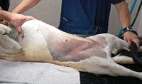
5. Widely clip the dorsally recumbent, anesthetized patient from the xiphoid cartilage to the pelvic brim, following the caudal edge of the rib cage to about halfway to the dorsum of the lumbar area (lateral to the third and fourth mammary glands) and straight across caudally where the cranial thigh meets the body. It is important to have a sterile field in the area where the ovarian tacking needles will be placed or in case the doctor needs to convert to an open procedure.
6. Ensure that the patient's bladder has been emptied.
During the procedure, the technician and veterinarian must ensure that the abdominal pressure does not exceed 15 mm Hg during the initial insertion of the Veress needle and primary and secondary cannulas since this pressure will impede the patient's respiration. Immediately after placement of the second cannula, reduce the intra-abdominal pressure to 8 to 10 mm Hg for the remainder of the procedure.
Tom McCabe, DVM
Northeast Veterinary Clinic
9405 Dyer St.
El Paso, TX 79924


 in general practice")


