Heartworm disease: What lies beneath
Dr. Stephen Jones takes us on a pictorial journey of the gross effects of heartworm infections in veterinary patients.


A look at the effects of heartworm disease in veterinary patients can help bring home the importance of keeping infections from spiraling out of control. (All photos courtesy of Dr. Stephen Jones)Let's start by evaluating our first impressions. Does this dog appear to have heartworm disease?


With a Grade V/VI holosystolic murmur, delayed capillary refill time, dyspnea, pulmonary crackles, weak femoral pulses, ascites and pitting edema, she certainly fits the bill.
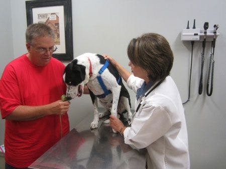
But what about this dog?


He's happy and healthy by every measurable clinical standard, but if you could see beneath the surface, you would know that he has heartworm disease, too. Veterinarians once defined a clinically normal dog like this as having heartworm infection but not heartworm disease. Let's evaluate a few more pictures as an illustration of how heartworm disease begins long before clinical signs develop and continues to progress as long as heartworms are present.
Heartworm infection begins with the bite of an infected mosquito like this irritating sucker.

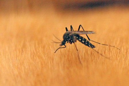
The infective larvae penetrate the dog's skin, spending between 67 and 125 days in the subcutaneous and somatic tissues of the body before the worms reach the young adult stage. At this point, the worms find their way into the pulmonary arteries, where they continue to mature.
If you could look inside the lungs of a normal, healthy dog prior to heartworm infection, here's what you'd see.

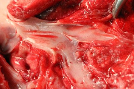

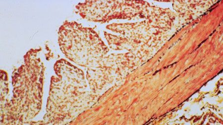
This image shows the distal pulmonary artery. The vascular walls are thin and the endothelial surface is smooth.
Contrast the appearance of a normal pulmonary artery with this image taken from a dog just 153 days (< six months) after experimental infection with heartworms.

Vascular endarteritis and proliferation within the distal pulmonary arteries are already apparent, in spite of the fact that these infections most likely would not be detectable with an antigen test.
Microscopically, we can see endarteritis depicted as villous proliferation of the endothelial surface and smooth muscle thickening within the vascular walls.


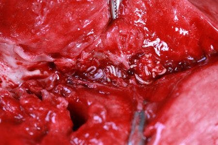
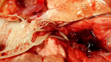
As the heartworms reach their full adult length of 10 to 12 inches, they begin to affect the larger, more proximal pulmonary arteries where the endarteritis progresses.

The greater the number of worms, and the longer the worms remain, the greater the likelihood that pathology will become significant.
As heartworm infection persists, the proliferation of the endothelial surface continues to worsen, and the vascular walls can become thickened and fibrotic.
These large-vessel changes, along with coinciding perivascular inflammation and smaller arterial and capillary obstruction, can dramatically affect blood flow. This leads to increases in pulmonary artery pressures and, ultimately, right-sided heart failure.
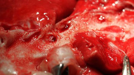
While adult heartworms can live up to five to seven years, their lifespan varies considerably. Some worms may never reach the adult stage, while others, as in this example, can die as young adult worms.

As part of the normal progression of heartworm disease, these dead worms collapse and are forced by the blood flow into the smaller distal branches of the pulmonary arteries.
Thromboembolic events ensue, often causing complete vascular obstruction and significant perivascular, peribronchiolar and alveolar disease. Symptomatically, the result can be coughing and, in more advanced cases, right-sided heart failure.

Keep in mind, the longer heartworm infection persists, the more this type of embolic event can occur, and the more likely clinical disease will develop.
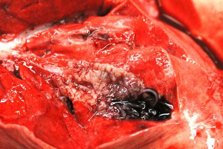
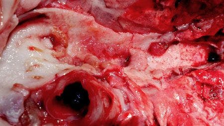
Dead heartworms aren't necessarily good heartworms. Rather than simply decomposing and being cleaned up by the dog's immune system, dead worms can leave behind chronic obstructive disease.

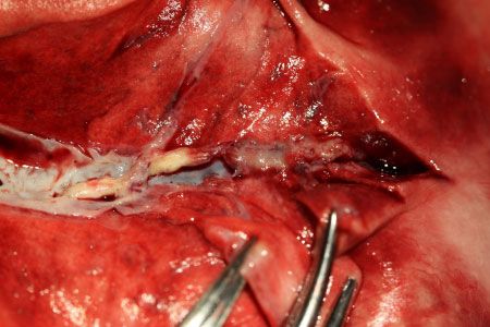
This image shows a dilated distal pulmonary artery with mummified adult worm remnants and intraluminal fibrosis causing permanent obstruction of blood flow.
Below we see mummified adult worm remnants in two small branches of a distal pulmonary artery.
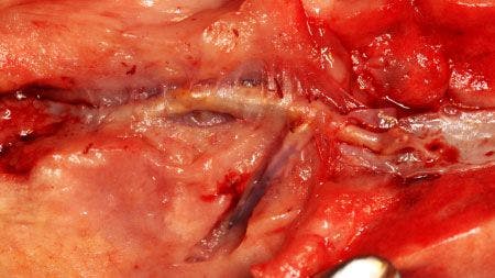
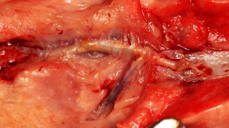
Obviously, changes such as this are permanent, leaving pets with irreversible disease.
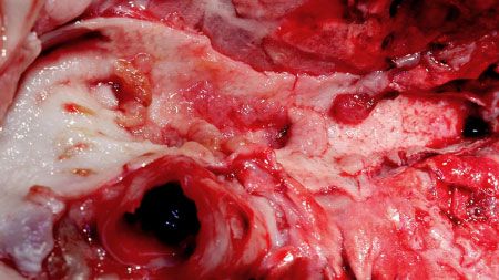
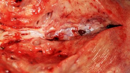
In this next image, we can see that the thickened vessel on the left ends abruptly as the more distal artery has completely become obstructed by fibrosis.

Take another look at this healthy distal pulmonary artery.
This is what we want for our patients, but it means being diligent about heartworm prevention. While there is some individual variation from one dog to another in how aggressively changes take place, all dogs with heartworm infection ultimately develop significant disease.
Remember: by the time a dog is diagnosed as heartworm-positive, heartworm disease has already begun.


Early diagnosis and early elimination of heartworms with adulticidal therapy give a dog the best chance of a healthy life, but heartworm infection has consequences, and damage can be life-long.
That's why the American Heartworm Society (AHS) recommends year-round heartworm prevention for all pets. For more information on the AHS guidelines, as well as tools to educate your staff and clients, check out heartwormsociety.org.
Dr. Stephen Jones is a board member of the AHS and immediate past president. He is a practice partner at Lakeside Animal Hospital in Moncks Corner, South Carolina.
Why all of the photos of heartworms? Dr. Jones says seeing has long been an element of believing when it comes to heartworm education-it's why countless veterinary practices display heartworms in a jar for client viewing. Seeing how heartworms damage the lungs and arteries of patients also helps veterinarians understand heartworm disease in ways that just reading about it can't do. That's why he goes to great lengths to document the disease damage he sees. His goal? To help veterinarians better understand the complex disease they both prevent and treat.