How to perform a scrotal urethrostomy and stop the stone cycle
Male dogs suffering from recurrent urethral blockage due to urinary calculi may benefit from this procedure that permanently diverts urine flow.
Male dogs suffering from recurrent urethral blockage due to urinary calculi may benefit from this procedure that permanently diverts urine flow.
Next >


Scrotal urethrostomy is a permanent urinary diversion procedure performed in dogs that have chronic or recurrent urethral blockage due to urinary calculi. Urethral calculi in male dogs may cause partial or total urethral obstruction by lodging in the urethra, usually at the base of the os penis. Less common indications for urethrostomy include urethral stricture from previous calculus injury or surgery, penile or preputial neoplasia, or severe trauma that necessitates penile and preputial amputation.
Scrotal urethrostomy requires scrotal ablation and neutering of intact male dogs. Complications of urethrostomy in dogs include hemorrhage from the urethra during the immediate postoperative period and an increased risk of urinary tract infection long term. Stricture of the surgical site is uncommon because of the width of the urethra at this level. Hemorrhage and stricture are both minimized by accurate apposition of the urethral mucosa to the skin.
Preoperative assessment and preparation
Before the surgery, perform appropriate blood work to ensure the patient’s stability, giving special attention to renal and electrolyte values. Perform abdominal and perineal radiography or ultrasonography to assess the entire urinary tract for calculi. After anesthesia is induced, position the dog in dorsal recumbency and prepare the caudal abdomen, including the prepuce and scrotum, for surgery by clipping and performing appropriate aseptic preparation.
If the patient is urethrally obstructed, calculi can be flushed back into the urinary bladder via retropulsion involving careful catheterization and fluid lavage while the patient is anesthetized but before starting the urethrostomy procedure. If cystic calculi are present, perform a cystotomy before starting the urethrostomy procedure to remove all bladder calculi. If possible, place an indwelling urethral catheter, which is to be maintained during the urethrostomy. Perform the urethrostomy before the bladder and abdominal walls are closed to allow for normograde and retrograde urethral catheter passage and lavage to ensure urethral patency.
How to perform a scrotal urethrostomy: A step-by-step guide

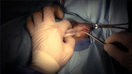
Step 1:
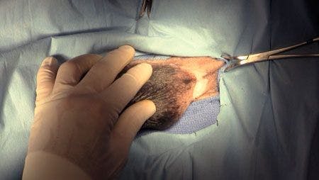
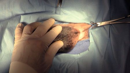
In intact dogs, make an elliptical skin incision near the base of the scrotum, preserving about 1 cm of scrotal skin on each side. (This dog’s head is to the left.) Excise the scrotal skin, and neuter the dog with an open or closed technique. In neutered dogs, excise the scrotal remnant or make a midline skin incision over the caudal penile body, extending caudally until the urethra starts to curve dorsally.


Step 2:
Incise subcutaneous fat to expose the ventral penile body and the retractor penis muscle attached to the penis. Bluntly elevate the retractor penis muscle, and displace it laterally to either side, exposing the urethra. The urethra is purple and located on the ventral midline of the penis.


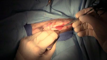
Step 3:
Use the thumb and index finger of your nondominant hand to elevate the penis from the incision before incising the urethra, which results in decreased hemorrhage from the incised corpus spongiosum tissue surrounding the urethra.

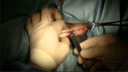
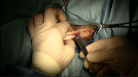
Step 4:
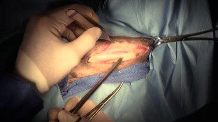
Use a No. 11 or No. 15 scalpel blade to incise the urethra on the midline over the previously placed urethral catheter. After you enter the urethral lumen, use tenotomy or iris scissors to extend the mucosal incision to a total length of 3 or 4 cm (see photo above).
Digitally elevate the urethra to decrease hemorrhage. Suctioning may improve the visibility of the urethral lumen and, especially, the urethral mucosal edge, which tends to retract away from the penile body (see photo directly above).
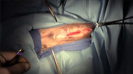


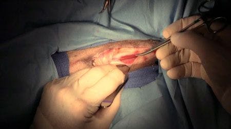
Step 5:
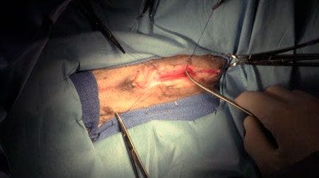
Using 4-0 polypropylene suture on a tapered needle, suture the cranial and caudal aspects of the urethral incision to the skin to establish the boundary of the urethrostomy. Careful suturing of the urethral mucosa to the skin is emphasized. Initially, stay sutures are placed to provide tension on the ends of the incision and will be tied later (see photos above).
The sequence of urethrostomy suturing is as follows: First incorporate a 2- or 3-mm purchase of urethral mucosa only, then a small purchase of the tunica albuginea (white fibrous-appearing tissue of the penis), and, finally, a small, angled purchase of the scrotal skin with the needle. The skin purchase engages the dermis and epidermis only. Taking the purchases separately minimizes mucosal trauma. This sequence of needle passage produces compression of the corpus spongiosum tissue and reduces postoperative hemorrhage from incised tissue.
After the corners have been sutured, place a simple continuous suture pattern between the caudal and cranial sutures on each side. End the continuous pattern by tying it to the tags of previously placed interrupted or stay sutures.
Modifications of this technique include the use of absorbable suture, such as poliglecaprone 25 or glycomer 631. Interrupted sutures may be placed to construct the urethrostomy stoma, with the medial suture tag being cut short.
Step 6:
At the conclusion of surgery, an 8- to 10-F red rubber catheter (not pictured here) should be able to be easily passed from the urethral stoma to the urinary bladder.
Watch of a video of Dr. Waldron performing this procedure.
Postoperative care
Urethral catheterization is not maintained after surgery. Place a self-restraint collar, and advise owners to keep it on the patient for one week. Occasional dripping of blood concurrent with or at the end of urination is common for the first three to five days after surgery. Nonabsorbable sutures are removed two weeks after surgery with the patient sedated, and absorbable sutures will fall out spontaneously.
And see even more step-by-step surgical videos by Dr. Waldron at dvm360.com/Waldron.


 in general practice")


