How to perform a surgical hepatic biopsy
In patients with known or suspected liver disease, obtaining a biopsy sample is often indicated. This article features guidance on when and how to perform a surgical hepatic biopsy.
In patients with known or suspected liver disease, obtaining a biopsy sample is often indicated. Liver samples may be obtained by a variety of techniques.1-4 Surgical biopsy allows the whole liver to be visually inspected and palpated, providing an ideal opportunity to obtain biopsy samples of focal lesions for histologic examination, culture and antimicrobial sensitivity testing, and metal analysis. Surgical biopsy also provides a larger tissue sample for a more complete histologic review and allows the veterinarian to examine the biopsy site for adequate hemostasis.2 This article features guidance on when and how to perform a surgical hepatic biopsy.
INDICATIONS
The indications for performing a liver biopsy include substantially or persistently increased liver enzyme activities or serum bile acid concentrations, hepatic hyperbilirubinemia, generalized changes in hepatic ultrasonographic echogenicity, unexplained hepatomegaly, or the presence of solitary or multiple focal lesions within the hepatic parenchyma.1 Liver biopsy is particularly important when the results of biochemical testing and advanced imaging modalities such as ultrasonography or scintigraphy are not adequate to establish a diagnosis. Additionally, histologic information can be combined with an already determined diagnosis to tailor specific treatment protocols, evaluate the response to therapy, and determine the prognosis. Specific indications for a surgical biopsy include microhepatia, a hepatic abscess or cyst, or a lesion that is difficult to localize on ultrasonographic examination.1 In addition, excisional biopsy of a pedunculated or solitary mass may provide treatment opportunities.
SELECTING AND COMPARING LIVER BIOPSY METHODS
Liver samples may be obtained percutaneously by blind or ultrasound-guided biopsy or surgically by laparoscopy or celiotomy.1-4 Selecting the appropriate method is determined by the size of the liver, the type and size of the lesion, and the patient's size and overall health.1,2
Biopsy samples from unstable patients or from patients with severe coagulopathy are best obtained by percutaneous means such as fine-needle aspiration or Tru-Cut biopsy, particularly if diffuse disease is suspected.1 However, both of these methods are significantly inferior to wedge biopsy, potentially affecting an accurate diagnosis.5-8 Cytologic examination of samples obtained by fine-needle aspiration cannot provide information about tissue architecture and may be nondiagnostic for lesions that exfoliate poorly. The correlation between cytologic examination of fine-needle liver aspirates and histologic examination of hepatic biopsy samples is poor; diagnostic correlation of samples was 30% in dogs and 51% in cats in one study and 29% in multiple species in another study.5,6 In fact, cytologic and histologic examinations have a significantly lower correlation in hepatic tissue than all other tissues studied, including cutaneous, subcutaneous, nasal, osseous, lymphatic, splenic, gastrointestinal, cerebral, and ocular tissues.6
Sample size and quality are also variable when ultrasound-guided tissue core biopsy samples are obtained. In one study, only 77% of intended liver biopsies retrieved hepatic tissue, and 22% of the samples were less than 2 mm long and lacked appropriate lobular architecture.7 Thirteen percent of the samples were completely devoid of tissue, and another 10% contained skeletal muscle, blood, or small intestine. Only 60% of samples obtained by Tru-Cut biopsy were of diagnostic quality.7
In another study, samples obtained with 18-ga spring-triggered biopsy needles using ultrasound guidance or direct observation during laparotomy or immediately postmortem had one-third to one-fourth of the median surface area of those obtained with wedge biopsy.8 Because of this small sample size, a diagnosis based on needle biopsy correlated with that of wedge biopsy in less than half the patients. Additionally, the severity of pathology was graded as significantly less in needle biopsy samples for most morphologic parameters, with the exception of inflammation, which was graded more severe as compared with wedge samples. Smaller needle biopsy samples were unable to achieve the necessary number of portal triads per sample to provide an accurate diagnosis of portal triad diseases such as portal venous hypoplasia or portal atresia and were subject to misinterpretation by evaluators.8 Laparoscopic biopsy allows better visualization of the site compared with ultrasonography but may provide less tissue volume compared with a surgical wedge biopsy.
PERIOPERATIVE CONSIDERATIONS
All patients should undergo presurgical diagnostic tests (complete blood count, platelet count, serum chemistry profile, coagulation tests, urinalysis) to determine which perioperative treatments should be administered. For example, animals with liver disease may present with vomiting that could result in dehydration and electrolyte abnormalities. Preoperative potassium or magnesium deficiencies may cause ileus, arrhythmias, and other complications during or after surgery, so they are best corrected before anesthesia. Animals with severe liver disease or sepsis may have prolonged clotting times, requiring crossmatching and transfusion before surgery.2,3 Animals suspected of having neoplasia should undergo staging of their disease, including thoracic radiography and abdominal ultrasonography. Aspirates obtained during ultrasonography may provide a diagnosis in some patients, obviating the need for surgery.
If possible, fast patients for 12 hours before surgery. Preoperatively, administer fluids and analgesics. Give hypoalbuminemic patients colloidal fluids such as hetastarch or plasma to provide oncotic pressure support. Choose premedicants and dosages with care since metabolism of some drugs may be delayed when liver dysfunction is present. We commonly use an opiate combined with a benzodiazepine in dogs and cats or a combination of ketamine and diazepam in cats. If acepromazine is administered as a sedative, the total dose should not exceed 0.25 mg. Induction can be performed with intravenous propofol or by mask induction with isoflurane. Clip patients undergoing liver biopsy to the midsternal level, and clip patients receiving feeding tubes farther laterally on the abdomen. All patients should have an appropriately sized endotracheal tube with an inflated cuff. During anesthesia, continuous-rate infusions of fentanyl can be administered to reduce gas anesthetic requirements.9,10 These infusions can be continued postoperatively to provide analgesia.
Ideally, respiratory and cardiac function and oxygenation should be monitored during anesthesia with a capnograph, electrocardiograph, arterial blood pressure monitor, and pulse oximeter. Maintaining intraoperative blood pressure with fluids and oncotic support is critical in patients with liver disease since reduced hepatic perfusion can have deleterious effects on postoperative liver function. Forced-air heating blankets (e.g. Bair Hugger—Arizant Healthcare) can be used to keep patients warm during the procedure.
Prophylactic antibiotics are usually unnecessary in patients undergoing liver biopsy. Be prepared to take culture samples during the procedure and to place feeding tubes in some patients. Cautery should be available intraoperatively for patients with coagulopathies. Count the sponges and laparotomy pads before the abdomen is opened and again before it is closed to prevent iatrogenic peritoneal foreign bodies. Suction all fluid from the abdominal cavity before closure.
Postoperative treatments, complications, and prognosis vary depending on the underlying disease process and the patient's condition. Most animals continue to receive fluid support and analgesics after surgery. In patients that are not vomiting, small amounts of food and water can be offered within eight hours of the procedure.
OPEN SURGICAL BIOPSY TECHNIQUE
Make a ventral midline abdominal incision. The incision should extend cranially to the level of the xiphoid to improve exposure of the liver, particularly if it is small. Liver exposure can be improved by removing the falciform fat and by carefully incising the triangular ligaments or by placing moistened laparotomy pads between the liver and the diaphragm.2,3 Examine the entire liver visually and by gentle palpation for nodules, cavitations, and other abnormalities.
Obtain samples at the junction of normal and diseased tissue to ensure that both abnormal and normal hepatocellular structures are included.11 If the liver is diffusely affected, obtain biopsy samples from the most accessible location, usually the liver margin. In conditions in which lesions are distributed irregularly, obtain samples from multiple lobes to increase the likelihood of obtaining a diagnostic sample. Although affected liver margins typically suggest parenchymal disease, their greater distance from hepatic blood supply may predispose them to fibrosis, obscuring the underlying pathology. Misdiagnosis of hepatic fibrosis can be avoided by taking larger or multiple samples.2,12

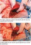
Figures 1,2
Peripheral or diffuse lesions
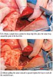
The guillotine method is used to sample the hepatic margin of pointed or sharp-edged lobes. Form a loop with 3-0 absorbable monofilament suture, using a single throw. Drop the suture loop over the point of the lobe, settling the suture into a natural fissure or in notches made by Kelly forceps (Figure 1). Tighten the suture completely so that it crushes all of the tissue within the loop, leaving the piece of tissue attached only by vessels and ducts (Figure 2). While tightening, do not pull the suture outward or upward, as this may sever the vessels and ducts and accidentally remove the ligature with the sample. A second throw can be placed to form a knot but is not necessary for hemostasis and can increase the risk of tearing the vessels. Transect tissue 2 to 3 mm distal to the suture from the lobe with Metzenbaum scissors or a scalpel blade. If you use a blade, place a finger under the piece of liver to be removed, and press the blade gently and firmly through the tissue toward the finger (Figures 3 & 4). If you press slowly but firmly with the flat portion of the cutting edge, you will easily cut through the liver tissue but will not damage your gloves. Trim the suture ligature short. To avoid iatrogenic specimen artifacts, do not use tissue forceps to handle the tissue sample.2,3 Excessive bleeding can be controlled with pressure, ligation, or cautery.2,4


Figures 3,4
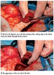
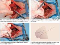
For marginal lesions on rounded liver lobes, place two parallel, full-thickness guillotine sutures perpendicular to the liver margin around the proposed site by using absorbable suture with a swaged needle (Figures 5-8). Leave the ends of the second suture knot long, and then pass one end of the suture around the base of the tissue pedicle and tie it back to the other end (Figures 9 & 10). This will crush the tissues across the base of the pedicle, resulting in hemostasis along all three sides of the tissue sample. Tighten the sutures completely so that they cut through the tissues, and remove the tissue within the suture box with scissors (Figures 11 & 12).


Figures 5,6,7,8
Central lesions
Samples from nonmarginal liver can be obtained with a skin biopsy punch, Tru-Cut biopsy needle, or laparoscopic clamshell biopsy instrument.12 To avoid nicking the large, dorsally located hepatic veins, take the sample, preferably, from the ventral hepatic surface, and do not let it exceed half the thickness of the lobe.

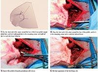

Figures 9,10,11,12
When taking biopsy samples of the liver with clamshell forceps, place the instrument against the site of interest with the jaws open. Insert the forceps into the parenchyma to the level of the angle of the jaws, and close the jaws. After a few seconds of crushing, twist the instrument until a free piece of tissue is removed. Pulling the forceps straight out of the liver tends to cause more hemorrhage than twisting.12
Hemorrhage from punch, Tru-Cut, or clamshell biopsy sites can be controlled by a variety of methods. Absorbable gelatin foam (Gelfoam—Pharmacia & Upjohn) can be inserted into the defect, a mattress suture of 3-0 absorbable material can be placed gently around the defect, or an omental flap can be sutured over the defect. Alternatively, pressure can be applied to the site, or the site can be cauterized by using a low setting.4,13 Thoroughly lavage the abdomen with a balanced electrolyte solution in patients with bile leakage, excessive hemorrhage, or infected or necrotic lesions.2
COMPLICATIONS
Complications after liver biopsy are uncommon but may include bile peritonitis, hemorrhage, and sepsis. The risk of complications is greater in patients with coagulopathies and thrombocytopenia.14,15 Major complication rates during hepatic biopsies have been reported to be as high as 22% and 50% in dogs and cats that are thrombocytopenic.14 Many patients with liver disease are debilitated from hypoalbuminemia and compromised liver function, increasing the risk of potential complications with anesthesia and surgery such as hypotension and altered metabolism of anesthetic and analgesic drugs.
Diana Burger, BS
Amie Carrier, BS
Karen M. Tobias, DVM, MS, DACVS
Department of Small Animal Clinical Sciences
College of Veterinary Medicine
The University of Tennessee
Knoxville, TN 37996-4544
REFERENCES
1. Day DG. Indications and techniques for liver biopsy. In: Textbook of veterinary internal medicine. 5th ed. Philadelphia, Pa: WB Saunders Co, 2000;1294-1298.
2. Martin RA, Lanz OI, Tobias KM. Liver and biliary system. In: Small animal surgery. 3rd ed. Philadelphia, Pa: WB Saunders Co, 2003;713-717.
3. Fossum TW. Surgery of the liver. In: Small animal surgery. 2nd ed. St. Louis, Mo: Mosby, 2002;450-457.
4. Harvey CE, Newton CD, Schwartz A. The liver and biliary tract, spleen, and pancreas. In: Small animal surgery. Philadelphia, Pa: JB Lippincott Co, 1990;407-411.
5. Wang KY, Panciera DL, Al-Rukibat RK, et al. Accuracy of ultrasound-guided fine-needle aspiration of the liver and cytologic findings in dogs and cats: 97 cases (1990-2000). J Am Vet Med Assoc 2004;224:75-78.
6. Cohen M, Bohling MW, Wright JC, et al. Evaluation of sensitivity and specificity of cytologic examination: 269 cases (1999-2000). J Am Vet Med Assoc 2003;222:964-967.
7. de Rycke LMJH, van Bree HJJ, Simoens PJM. Ultrasound-guided tissue-core biopsy of liver, spleen and kidney in normal dogs. Vet Radiol Ultrasound 1999;40:294-299.
8. Cole TL, Center SA, Flood SN, et al. Diagnostic comparison of needle and wedge biopsy specimens of the liver in dogs and cats. J Am Vet Med Assoc 2002;220:1483-1490.
9. Valverde A, Doherty TJ, Hernandez J, et al. Effect of lidocaine on the minimum alveolar concentration of isoflurane in dogs. Vet Anaesth Analg 2004;31:264-271.
10. Hellyer PW, Mama KR, Shafford HL, et al. Effects of diazepam and flumazenil on minimum alveolar concentrations for dogs anesthetized with isoflurane or a combination of isoflurane and fentanyl. Am J Vet Res 2001;62:555-560.
11. Garner MM, Raymond JT, Toshkov I, et al. Hepatocellular carcinoma in black-tailed prairie dogs (Cynomys ludivicianus): tumor morphology and immunohistochemistry for hepadnavirus core and surface antigens. Vet Pathol 2004;41:353-361.
12. Richter KP. Laparoscopy in dogs and cats. Vet Clin North Am Small Anim Pract 2001;31:707-727.
13. Kim EH, Kopecky KK, Cummings OW, et al. Electrocautery of the tract after needle biopsy of the liver to reduce blood loss. Invest Radiol 1993;28:228-230.
14. Bigge LA, Brown DJ, Penninck DG. Correlation between coagulation profile findings and bleeding complications after ultrasound-guided biopsies: 434 cases (1993-1996). J Am Anim Hosp Assoc 2001;37:228-233.
15. Weber JC, Navarra G, Jiao LR, et al. New technique for liver resection using heat coagulative necrosis. Ann Surg 2002;236:560-563.


 in general practice")


