Image Quiz: What do you see in this cytology slide?
This skin impression cytology sample is from a 5-year-old, mixed-breed, spayed female dog.
The patient has multifocal cutaneous lesions on the dorsal and lateral trunk, with papules, epidermal collarettes and mild crusting. What is your primary differential?

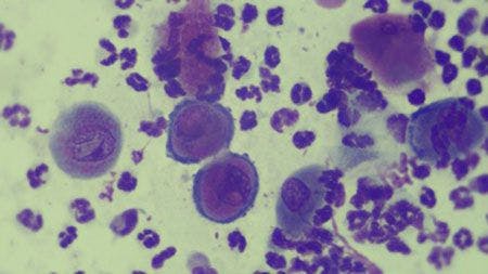
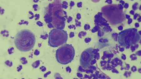
Images courtesy of Dr. Chris Reeder.
A. Mast cell tumor
B. Pemphigus foliaceus
C. Discoid lupus erythematosus
D. Cutaneous lymphoma
Click to the next page for the answer.
The correct answer is B: Pemphigus foliaceus

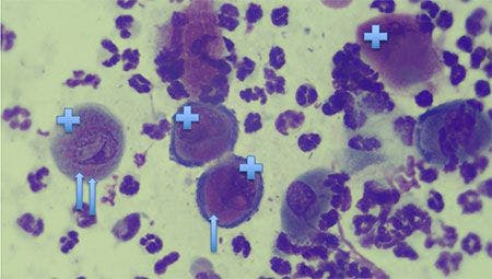
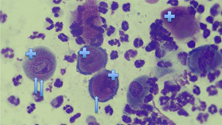
The large round cells are acantholytic cells (+) with keratohyaline granules (arrows; pink intracellular blobs), which define these as keratinocytes and not macrophages or lymphoid cells. Numerous neutrophils are present in the sample as well, although no bacteria or yeast can be seen. Pemphigus foliaceus was confirmed via skin punch biopsies. This is the most common autoimmune skin disease seen in dogs. Its three main causes are:
hereditary in Akitas and chow chows
chronic skin disease (e.g. atopic dermatitis)
medication-induced (e.g. antibiotics, flea/tick products).
Treatments for pemphigus foliaceus vary depending on the severity of the condition, the health of the dog and the dog's previous medication history. Most commonly, oral modified cyclosporine, prednisone and/or azathioprine are initial courses. If you are not familiar with treatment of pemphigus, it's best to consult with your local/regional veterinary dermatologist.
Dr. Chris Reeder practices at BluePearl Veterinary Partners in Franklin, Tennessee. Dr. Reeder's special interests include ear disease and immune-mediated skin diseases. He is an avid hobby farmer raising a large number of critters.