Just Ask the Expert: Can a smoky sclera be normal?
Dr. Juliet Gionfriddo answers this reader query about a discolored sclera.
Dr. Gionfriddo welcomes ophthalmology questions from veterinarians and veterinary technicians.
Click here to submit your question, or send an e-mail to vm@advanstar.com with the subject line "Ophthalmology questions."
One of my patients is an 8-month-old English bulldog with a unilateral smoky-colored sclera (see photo, left). I have never seen this before and am interested in your opinion.


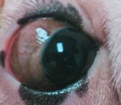
A. The bulldog in your picture has more extensive scleral and episcleral pigmentation than we usually see in dogs, but it is normal. In dogs, the color of the sclera depends on the thickness of its stroma, the amount of pigment it contains, and the amount of fat in its outer boundary. The sclera is thicker near the limbus and thins toward the equator of the globe, making it appear whiter at the limbus in some individuals. However, it is common in dogs to have a large amount of limbal melanin that makes the limbus appear dark-brown or black. This pigment sometimes extends posteriorly for a few millimeters either around the entire limbus or may be particularly prominent laterally (Figure 1). In the case of your bulldog, the melanin is scattered lightly throughout the sclera, making it appear gray, and the pigment extends over the entire sclera rather than being focal. This may be related to the dog's coat color.

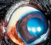
1. A dog with temporal limbal pigmentation, which is a normal finding.
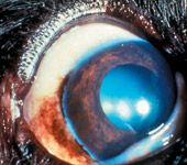
This gray-appearing scleral pigmentation must be differentiated from a very thin sclera, which appears dark-blue because of the dark uveal tract that can be seen through the thin sclera. The most common reason for scleral thinning is the presence of a staphyloma, which is a scleral defect that is lined with uveal tissue. A staphyloma is usually localized to a portion of the eye and does not involve the entire sclera, unlike the pigment in this bulldog. In addition, staphylomas often bulge from the ocular surface (Figure 2). They can be congenital because of developmental defects in scleral formation or traumatic. If they are traumatic in origin, staphylomas should be corrected surgically. Large, congenital staphylomas also may be surgically corrected, but if they are small, they are often left alone.


2. A young dog with a congenital staphyloma of the temporal portion of the globe.
Juliet R. Gionfriddo, DVM, MS, DACVO
Department of Clinical Sciences
College of Veterinary Medicine and Biomedical Sciences
Colorado State University
Fort Collins, CO 80523


")

")


