Just Ask the Expert: How should I approach a discolored tooth?
Our dentistry expert Dr. Daniel T. Carmichael answers this reader question.
Dr. Carmichael welcomes dentistry questions from veterinarians and veterinary technicians.
Click here to submit your question, or send an e-mail to vm@advanstar.com with the subject line "Dentistry questions."
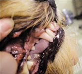
One of my patients is a 9-year-old Norwich terrier with an acute onset of intrinsic discoloration of its maxillary right canine (see photo). Do all cases such as this require root canal therapy? What is the prognosis? Would radiographs be of value?

Craig A. Nausley, DVM Briarcrest Veterinary Care Center Tucson, Ariz.
Think of this discoloration, which often changes color (pink to purple to gray) over time, as signifying a dead tooth.1 More specifically, some event has caused the dental pulp to become inflamed (pulpitis) and subsequently undergo necrosis. The tooth discoloration is pigment from red blood cell breakdown that has percolated into the dentin tubules. Inside the pulp chamber, the necrotic pulp contains various inflammatory mediators. They can seep out through the apical delta, leading to periapical periodontitis and other adverse sequelae.

Daniel T. Carmichael, DVM, DAVDC
RADIOGRAPHY IS VITAL
The first step in the diagnostic workup of a discolored tooth is to obtain an intraoral radiograph. The two most important questions to answer when evaluating the radiograph are 1) Is there pathology in the apical regions? and 2) What is the relative thickness of the dentin walls?
If there is any evidence of periapical pathology, such as periapical radiolucency or apical resorption, the tooth needs treatment. If the discolored tooth has thin dentin walls, this indicates that the tooth was immature when the damage occurred. Thin dentin walls are associated with a robust amount of necrotic pulp tissue, and this situation eventually causes problems if left untreated. Another indication for treatment would be if the history suggests oral pain associated with the discolored tooth.
When treatment is indicated, the options are to extract the tooth, or, ideally, perform root canal treatment. With treatment, the prognosis is generally excellent.
TREATMENT VS. MONITORING
Experts disagree on whether treatment is indicated when a discolored tooth has mature dentin walls and no radiographic evidence of periapical pathology. Some experts argue that any dead tooth has a chance of becoming a problem, so all discolored teeth should be treated. The opposing view assumes that in the mature tooth the body is able to wall off the necrosed pulp and essentially entomb the inflammation where it can do no harm. My personal opinion is that this subcategory of pulpal necrosis may be addressed by yearly radiographic monitoring rather than endodontics or extraction.

Key points
So most importantly, radiographs are essential in the treatment planning for this tooth (see sidebar "Key points"). This case is an illustration of why every veterinary practice offering dental service needs to be equipped with dental radiography.
Daniel T. Carmichael, DVM, DAVDC
Veterinary Medical Center
76 Sunrise Highway
West Islip, NY 11795
REFERENCE
1. Hale FA. Localized intrinsic staining of teeth due to pulpitis and pulp necrosis in dogs. J Vet Dent 2001;18(1):14-20.