Just Ask the Expert: Teeth fused to the mandible
Q. How do you proceed when you have a cat anesthetized and find that a tooth (or teeth) you need to extract is fused to the mandible?
Dr. Carmichael welcomes dental questions from veterinarians and technicians.
To ask your question, e-mail: vm@advanstar.com
With the subject line: Dental Questions
Q. How do you proceed when you have a cat anesthetized and find that a tooth (or teeth) you need to extract is fused to the mandible?
A. First, take a dental radiograph! In fact, the answer to almost every question pertaining to veterinary dentistry is “First, take a dental radiograph.” The results of an intraoral dental radiographic examination coupled with the results of an intraoral examination are essential when making proper and rational treatment decisions.
If a cat's tooth appears to be fused to the mandible as you describe, you are probably dealing with a dental resorptive lesion (also known as feline odontoclastic resorptive lesions, or FORL). Two types of resorption have been described in cats: type 1 and type 2. These resorptive lesions differ in their radiographic appearance (See Figures A & B) and are treated differently. With type 1 lesions, inflammatory resorption has occurred, and the entire tooth and root need to be extracted. These teeth are not fused to the bone, although they can be difficult to extract without proper equipment and technique. With type 2 lesions, replacement resorption has occurred and the tooth does become fused to the bone, so a subgingival coronectomy (or crown amputation) can be performed.


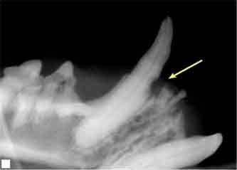
Figure A. A dental radiograph of the mandibular left canine tooth of a cat reveals a type 1 dental resorptive lesion (arrow). This tooth and its root should be extracted. The radiograph also reveals that the mandibular left third premolar has external crown resorption (type 1), a retained root of the mandibular left third incisor, and that the mandibular left first incisor is missing.
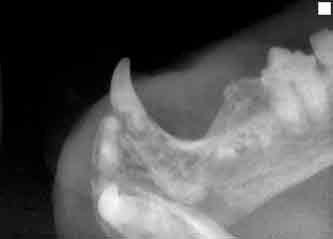

Figure B. A dental radiograph of the mandibular right canine tooth (in a different cat) reveals a type 2 dental resorptive lesion. Replacement resorption has occurred in almost the entire root, and the mandibular right third premolar also shows type 2 resorption of both roots. If the cat does not have gingivostomatitis, a subgingival coronectomy is the treatment of choice for both teeth.
REFERENCES
1. DuPont GA. Radiographic evaluation and treatment of feline dental resorptive lesions. Vet Clin North Am Small Anim Pract 2005;35(4):943-962.
2. Carmichael DT. How to detect and treat feline odontoclastic resorptive lesions. Vet Med 2005;100(2):102-110.
Daniel T. Carmichael, DVM, DAVDC
Veterinary Medical Center
75 Sunrise Highway
West Islip, NY 11795