Key gastrointestinal surgeries: Intestinal biopsy
You may be hesitant to perform a full-thickness incisional biopsy to obtain an intestinal tissue sample, but in many cases, this technique is preferred. In this article, we review when incisional biopsy is best and provide a simple step-by-step guide to the procedure to increase your confidence.
YOU MAY BE HESITANT to perform a full-thickness incisional biopsy to obtain an intestinal tissue sample, but in many cases, this technique is preferred. In this article, we review when incisional biopsy is best and provide a simple step-by-step guide to the procedure to increase your confidence. For general perioperative considerations when performing this procedure, including diagnostic testing, patient monitoring, and postoperative support, please see the symposium introduction.
INDICATIONS AND METHODS
Indications for intestinal biopsy include chronic diarrhea, vomiting, or weight loss; protein-losing enteropathy; or an intestinal mass.1 Biopsy samples can be obtained endoscopically, percutaneously by using ultrasound guidance, or surgically during laparotomy or laparoscopy.1-4
Many veterinarians prefer endoscopic biopsy because it is minimally invasive and permits direct visualization and sampling of the focal lesions. However, endoscopic biopsy samples can be routinely obtained only from the duodenum and colon, and lesions below the mucosa will be missed with this technique.4 For example, neoplastic and inflammatory lesions seen with lymphoma and feline infectious peritonitis, respectively, often do not extend into the mucosa.5,6 Dilated lymphatic vessels in dogs with intestinal lymphangiectasia may be limited to the mucosa-submucosa junction, making endoscopically obtained biopsy samples nondiagnostic.7 Lymphatic lesions can also be artifactually eliminated with iatrogenic collapse of the lacteals during sample retrieval with flexible biopsy forceps.7
Core biopsy samples of the intestines can be obtained percutaneously by using ultrasound guidance. An automated microcore biopsy (Biopty-Cut biopsy needle—C.R. Bard) requires a bowel wall thickness of at least 2 cm and is best used on infiltrative lesions.2 However, a diagnosis based on histologic examination of ultrasound-guided percutaneous biopsy samples is correct in only 69% of patients; accuracy varies with the lesion's location and underlying etiology.2
Surgical biopsy via celiotomy is performed in patients that require concurrent surgical procedures such as liver biopsy, mass resection, or feeding tube placement or in patients with intestinal lesions that cannot be reached endoscopically.1 It is also performed when samples from an endoscopic mucosal biopsy are not diagnostic.7 Obtaining a full-thickness, high-quality sample by surgical biopsy minimizes the difficulty of histologically interpreting samples from dogs with inflammatory bowel disease and other disorders.7,8
Surgical intestinal biopsy samples are usually obtained with scalpel blades or scissors but can also be taken with a Keyes biopsy punch. With this technique, the procedure duration, sample quality, and postoperative complication rate are similar to those of techniques in which scissors or scalpel blades are used.1

Figures 1,2
SURGICAL TECHNIQUE FOR INCISIONAL BIOPSY OF THE INTESTINES
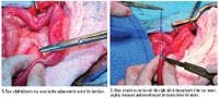
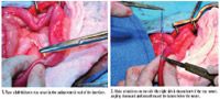
After thoroughly exploring the abdomen, isolate the affected area of intestine with moistened laparotomy pads. Multiple samples are usually taken, so include all potential biopsy sites and start with the least contaminated area (i.e. the small intestines). Milk the luminal contents away from the biopsy site. Place a full-thickness, 3-0 or 4-0 monofilament stay suture in the antimesenteric wall of the intestines perpendicular to the long axis of the intestines (Figure 1). Attach a hemostat to both ends of the stay suture, and elevate it away from the intestines. With a No. 15 scalpel blade, make an incision through the intestines on one side of the stay suture, angling downward and inward toward the lumen below the suture (Figure 2). Make a similar incision along the opposite side of the stay suture so that the biopsy sample is transected from its intestinal attachments (Figure 3). Drop the stay suture and attached intestinal sample into a formalin container; the pathologist will remove the suture once the sample is fixed.


Figures 3,4,5,6,7,8
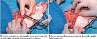
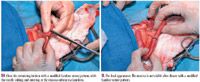
Close the enterotomy site (Figure 4) with a continuous or interrupted appositional pattern of 3-0 or 4-0 absorbable monofilament suture on a swaged, tapercut needle. To prevent mucosal eversion, perform a modified Gambee suture pattern.9 Insert the needle full thickness about 3 mm from the intestinal edge (Figure 5). Then back out the needle so the tip can exit at the mucosa-submucosa junction (Figures 6 & 7). Upward tension on the needle will facilitate proper placement since it causes the mucosa to roll downward over the needle tip and expose the white line of submucosa. Push the needle through the intestinal wall, and ready it for the next bite. Force the mucosa of the opposite side of the incision downward with the tip of the needle, and place a full-thickness bite, starting at the mucosa-submucosa junction (Figure 8). The first throw on the suture can either be a simple throw or a surgeon's throw and should be tightened enough to appose but not crush the intestinal wall. To prevent loosening, we use a surgeon's throw to begin the knot, particularly when tension is present. Add three or four additional throws to complete the knot (Figure 9). Small biopsy sites can be closed with three sutures: Place the initial appositional suture across the center of the incision, and add an additional suture to each side (Figures 10 & 11).
Figure 9
COMPLICATIONS
Complications are rare after intestinal biopsy. In one study, dehiscence of incisional biopsy sites was reported in 1.9% of dogs and was not correlated with systemic albumin concentrations.3 Dehiscence rates are similar for animals undergoing enterotomy for foreign body removal (2.6%).10 In another study, septic peritonitis and death secondary to biopsy site leakage were reported in one of 12 dogs undergoing intestinal biopsy.1

Figures 10,11
Laura Brandt, DVM
Karen M. Tobias, DVM, MS, DACVS
Department of Small Animal Clinical Sciences
College of Veterinary Medicine
The University of Tennessee
Knoxville, TN 37996-4544
REFERENCES
1. Keats MM, Weeren R, Greenlee P, et al. Investigation of Keyes skin biopsy instrument for intestinal biopsy versus a standard biopsy technique. J Am Anim Hosp Assoc 2004;40:405-410.
2. Crystal MA, Penninck DG, Matz ME, et al. Use of ultrasound-guided fine-needle aspiration biopsy and automated microcore biopsy for the diagnosis of gastrointestinal diseases in small animals. Vet Radiol Ultrasound 1993;34:438-444.
3. Harvey HJ. Complications of small intestinal biopsy in hypoalbuminemic dogs. Vet Surg 1990;19:289-292.
4. Hall EJ. Small intestinal disease—is endoscopic biopsy the answer? J Small Anim Pract 1994;35:408-414.
5. Couto CG, Rutgers HC, Sherding RG, et al. Gastrointestinal lymphoma in 20 dogs. A retrospective study. J Vet Intern Med 1989;3:73-78.
6. Harvey CJ, Lopez JW, Hendrick MJ. An uncommon intestinal manifestation of feline infectious peritonitis: 26 cases (1986-1993). J Am Vet Med Assoc 1996; 209:1117-1120.
7. Peterson PB, Willard MD. Protein-losing enteropathies. Vet Clin North Am Small Anim Pract 2003;33:1061-1082.
8. Willard MD, Jergens AE, Duncan RB, et al. Interobserver variation among histopathologic evaluations of intestinal tissues from dogs and cats. J Am Vet Med Assoc 2002;220:1177-1182.
9. Brown DC. Small intestines. In: Slatter D, ed. Textbook of small animal surgery. 3rd ed. Philadelphia, Pa: WB Saunders Co, 2003;644-664.
10. Weisman DL, Smeak DD, Birchard SJ, et al. Comparison of a continuous suture pattern with a simple interrupted pattern for enteric closure in dogs and cats: 83 cases (1991-1997). J Am Vet Med Assoc 1999;214:1507-1510.