Meningioma in a 9-year-old chocolate Lab: Anatomic pathology perspective
Dr. Jennie Jankovsky provides the anatomic pathology perspective on this challenging oncology case.


Dr. Jennie JankovskyCanine meningiomas, which arise from the meninges, are generally solitary masses located within the rostral and basilar regions of the calvarium or over the cerebral convexities (Figures 4 and 5), with other locations occasionally noted.1

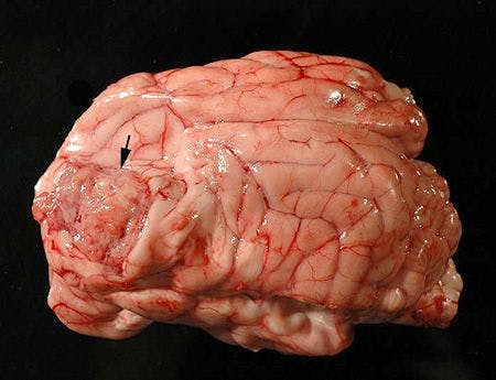

Figure 4. Meningioma (arrow) within the frontal lobe of another patient.


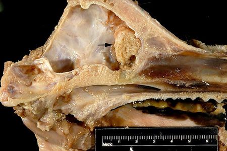
Figure 5. Meningioma (arrow) arising within the olfactory lobe region of another patient.
Intravertebral and orbital meningiomas are also infrequently reported (Figure 6).2,3 Grossly, these masses are firm, broad-based, well-circumscribed, ovoid to plaque-like, and have a smooth to granular surface. Metastasis is rarely reported to the lungs.4-6


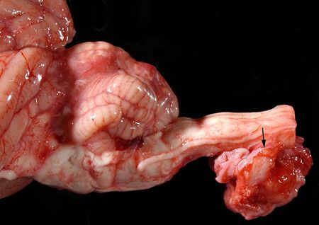
Figure 6. Meningioma (arrow) in the cervical spine of another patient.
The current World Health Organization (WHO) veterinary histological classification system divides meningiomas into benign and anaplastic (malignant) groups. The benign category is further characterized by histologic pattern, including meningothelial, fibrous, transitional, psammomatous, angiomatous, papillary and granular types.7 Meningothelial and transitional forms are most common in dogs.8,9
A grading system and additional histologic patterns are included within the WHO classification scheme for human meningiomas.10 Canine and human meningiomas are similar histologically, so evaluation of canine meningiomas with the human classification scheme allows greater prognostic capability.9 The WHO grading system for human meningiomas consists of three grades: benign (grade I), atypical (grade II), and malignant (grade III). Benign tumors lack histologic indicators of malignancy and include all histologic subtypes except clear cell, chordoid, papillary and rhabdoid. Atypical meningiomas are characterized by ≥ 4 mitoses/10 hpf, possible brain invasion, and three out of five of the following criteria:
• Cells arranged in sheets
• High nuclear to cytoplasmic ratio
• Hypercellularity
• Macronucleoli
• Necrosis
Additionally, clear cell and chordoid variants are included in the atypical category. Malignant meningiomas have ≥ 20 mitoses/10 hpf, features of malignancy similar to those described for atypical masses, and, if anaplastic, may resemble other neoplasms, such as carcinoma, sarcoma or melanosarcoma. The papillary and rhabdoid subtypes are included within the grade III category.10
Immunohistochemistry may be used to differentiate anaplastic meningiomas from other intracalvarial neoplasms. Meningiomas are immunoreactive to vimentin, CD34, E-cadherin, and doublecortin, with variable positivity for S-100, claudin-1, and neuron specific enolase (NSE).11-13 Neoplastic cells are not immunoreactive to synaptophysin, but are occasionally positive for cytokeratin and glial fibrillary acidic protein (GFAP), although often weakly or in a low percentage of cells.11,12
In the current case, histologic features were most consistent with a grade I meningothelial meningioma (Figures 7 and 8). Neoplastic cells had mild anisocytosis and anisokaryosis, with 3 mitoses/10 hpf. Necrosis and brain invasion were not present within the tissues examined.

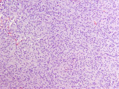
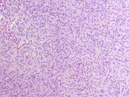
Figure 7. Meningothelial meningioma from the dog in this case. Epithelioid cells are arranged in sheets and loose whorls (hematoxylin and eosin stain; 100x).
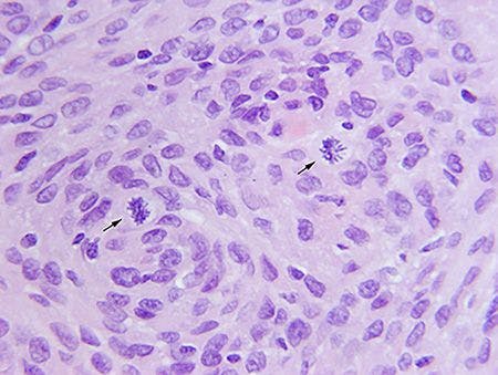
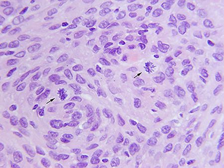
Figure 8. Meningothelial meningioma from the dog in this case. There are mitoses (arrows) (hematoxylin and eosin stain; 400x).
References
1. Motta L, Mandara MT, Skerritt GC. Canine and feline intracranial meningiomas: an updated review. Vet J 2012;192:153-165.
2. Mauldin EA, Deehr AJ, Hertzke D, et al. Canine orbital meningiomas: a review of 22 cases. Vet Ophthalmol 2000;3:11-16.
3. Petersen SA, Sturges BK, Dickinson PJ, et al. Canine intraspinal meningiomas: imaging features, histopathologic classification, and long-term outcome in 34 dogs. J Vet Intern Med 2008;22:946-953.
4. Dugan SJ, Schwarz PD, Roberts SM, et al. Primary optic nerve meningioma and pulmonary metastasis in a dog. J Am Anim Hosp Assoc 1993;29:11-16.
5. Schulman FY, Ribas JL, Carpenter JL, et al. Intracranial meningioma with pulmonary metastasis in three dogs. Vet Pathol 1992;29:196-202.
6. Pérez V, Vidal E, González N, et al. Orbital meningioma with a granular cell component in a dog, with extracranial metastasis. J Comp Pathol 2005;133,212-217.
7. Koestner A, Bilzer T, Fatzer R, et al. Tumors of meningothelial cells. In: World Health Organization histological classification of tumors of the nervous system of domestic animals. 2nd ed. Washington, DC: Armed Forces Institute of Pathology, 1999.
8. Patnaik AK, Kay WJ, Hurvitz AI. Intracranial meningioma: a comparative pathologic study of 28 dogs. Vet Pathol 1986;23:369-373.
9. Sturges BK, Dickinson PJ, Bollen AW, et al. Magnetic resonance imaging and histological classification of intracranial meningiomas in 112 dogs. J Vet Intern Med 2008;22:586-595.
10. Louis DN, Ongaki H, Wiestler OD, et al. Meningiomas. In: WHO classification of tumours of the central nervous system. 4th ed. Lyon, France: International Agency for Research on Cancer, 2007.
11. Barnhart KF, Wojcieszyn J, Storts RW. Immunohistochemical staining patterns of canine meningiomas and correlation with published immunophenotypes. Vet Pathol 2002;39:311-321.
12. Ramos-Vara JA, Miller MA, Gilbreath E, et al. Immunohistochemical detection of CD34, E-cadherin, claudin-1, glucose transporter 1, laminin, and protein gene product 9.5 in 28 canine and 8 feline meningiomas. Vet Pathol 2010;47:725-737.
13. Ide T, Uchida K, Suzuki K, et al. Expression of cell adhesion molecules and doublecortin in canine anaplastic meningiomas. Vet Pathol 2011;48:292-301.


")


