Meningioma in a 9-year-old chocolate Lab: Radiology perspective
Dr. Silke Hecht provides the radiology perspective on this challenging oncology case.


Dr. Silke HechtAlthough magnetic resonance imaging (MRI) is the modality of choice for brain imaging, computed tomography (CT) can provide valuable information about the presence, number and location of intracranial lesions, especially if they are large or contrast-enhancing. Whether neoplastic or not, intracranial masses can be subdivided based on location into intra-axial (arising from within the brain axis) and extra-axial.1-4 They can be further characterized by number, location, size, margination, signal intensity, homogeneity, contrast enhancement and concurrent imaging findings (e.g. ventriculomegaly, changes associated with the cranium or meninges, hemorrhage, mineralization, mass effect, edema, cystic or necrotic component).5-8
Extra-axial masses arise from tissues other than actual brain parenchyma (e.g. meningiomas and choroid plexus tumors). Many are located in the periphery of the brain and compress rather than invade brain parenchyma. Some are in a typical location (e.g. pituitary tumors originating from the pituitary fossa), and most are strongly contrast-enhancing. Concurrent changes to the skull (e.g. lysis or hyperostosis), broad-based contact of the lesion with meninges and skull forming an obtuse angle, and meningeal thickening and enhancement adjacent to a mass (“dural tail sign”) also suggest extra-axial origin.
Intra-axial masses originating from brain parenchyma may be completely surrounded by normal brain tissue, in which case determination of mass origin is straightforward. Peripherally located or very large masses may be in contact with overlying meninges, which may make a diagnosis challenging. An acute rather than obtuse angle of the mass with the meninges and absence of adjacent meningeal enhancement may be helpful in classifying a lesion as intra-axial in those cases. Finally, while the presence and degree of contrast enhancement of intra-axial masses is highly variable, absent or poor enhancement is most consistent with intra-axial origin and protection of the mass by the blood-brain barrier.
Imaging findings with intracranial meningioma include a round to ovoid or plaque-like smoothly marginated mass associated with the brain, typically in broad-based contact with underlying bone (except meningiomas in a ventricular location) (Figure 1).2-4,9-17


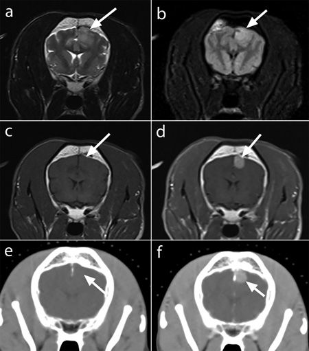
Figure 1. Meningioma in a 13-year-old West Highland white terrier (not the patient in this case). Transverse T2-weighted (a), T2-FLAIR (b), and T1-weighted MRI images before and after contrast administration (c, d); and transverse CT images before and after contrast medium administration (e, f). A focal peripheral well-circumscribed mass is associated with the left parietal lobe (arrows). The lesion is faintly hyperintense on T2-weighted and T2-FLAIR MRI images (a, b), isointense to brain parenchyma on the T1-weighted MRI image (c), isoattenuating to brain parenchyma on the pre contrast CT image (e), and strongly homogeneously contrast enhancing on both post contrast MRI and CT images (d, f).
They are usually single, although multiple tumors are possible.9,15,18,19 On CT images, meningiomas are typically isoattenuating or hyperattenuating (Figures 1e and 1f). On MRI, meningiomas are usually hypointense to isointense on T1-weighted images (Figures 1c and 2d) and hyperintense on T2-weighted/T2-FLAIR images (Figures 1a, 1b, 2a, 2b, and 3a).


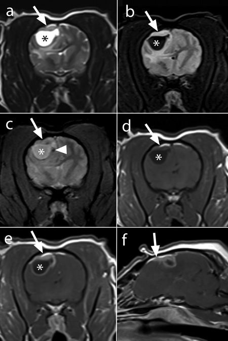
Figure 2. Cystic meningioma in a 13-year-old Dachshund (not the patient in this case). Transverse T2-weighted (a), T2-FLAIR (b), T2*-weighted (c), and T1-weighted MR images before and after contrast administration (d, e) and a sagittal T1-weighted MRI image after contrast medium administration (f). A peripheral plaque-like mass is associated with the right frontal and parietal lobes (a-e, arrows). This mass is mildly hyperintense to normal brain parenchyma on T2-weighted (a) and T2-FLAIR (b) images, isointense on T2*-weighted (c) and T1-weighted (d) images, and strongly homogeneously contrast enhancing (e). Associated with the ventral aspect of the mass is a large cyst, which is isointense to cerebrospinal fluid on all sequences (*). This cystic component is ring enhancing (e). Diffuse perilesional T2 and T2-FLAIR hyperintensity (a, b) is consistent with vasogenic edema, which results in a mass effect and midline shift toward the left. A focal punctate hypointense focus/susceptibility artifact seen on the T2*-weighted image (c; arrowhead) is consistent with focal mineralization or, less likely, focal hemorrhage. Thickened contrast enhancing meninges ("dural tail sign") extend peripherally from the mass (f; arrow).


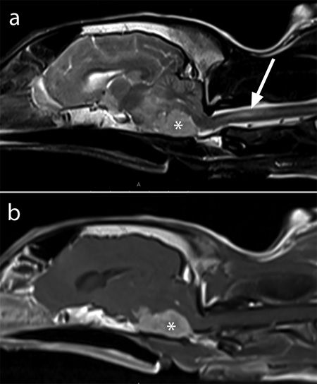
Figure 3. Caudal fossa meningioma in an 11-year-old mixed-breed dog (not the patient in this case). Sagittal T2-weighted (a) and post contrast T1-weighted (b) MRI images. A large T2 hyperintense and strongly homogeneously contrast-enhancing mass is associated with the caudal ventral cranial vault (*), which results in severe displacement and compression of the adjacent brainstem and cerebellum. A tubular T2 hyperintensity in the cervical spinal cord (a; arrow) is consistent with syringomyelia.
Strong contrast enhancement is seen with both imaging modalities, with homogeneous, heterogeneous or ring enhancement patterns possible (Figures 1d, 1f, 2e, 2f, and 3b).1-3,6,9-13,17,20-22 Concurrent common findings include brain edema (Figures 2a and 2b), mass effect3,5,12,15 and a “dural tail sign” (thickening and enhancement of the dura adjacent to the mass) (Figure 2f).15,23 Less common findings include mineralization or hemorrhage associated with the mass causing hyperattenuating areas on CT and T2 hypointense foci and/or susceptibility artifact (signal void) on T2*-weighted MRI images, respectively (Figure 2c).4,13,15,24,25 Additional findings may include bone changes adjacent to the tumor including hyperostosis, pressure atrophy or tumor invasion of bone (more common in cats),13,15,26,27 single or multiple tumor-associated cystlike changes (more common in dogs and predominantly seen in the rostral fossa) (Figures 2a-2e),13,15,28-31 brain herniation and cervical syringomyelia (Figure 3a), especially secondary to tumors in the caudal fossa.32-34
Other tumor types that may affect the meninges and resemble meningiomas include disseminated histiocytic sarcoma, lymphoma, granular cell tumors and metastatic disease (meningeal carcinomatosis).35-40
References
1. Kraft SL, Gavin PR. Intracranial neoplasia. Clin Tech Small Anim Pract 1999;14:112-123.
2. Kraft SL, Gavin PR, DeHaan C, et al. Retrospective review of 50 canine intracranial tumors evaluated by magnetic resonance imaging. J Vet Intern Med 1997;11:218-225.
3. Thomas WB, Wheeler SJ, Kramer R, et al. Magnetic resonance imaging features of primary brain tumors in dogs. Vet Radiol Ultrasound 1996;37:20-27.
4. Hecht S. Brain. In: Schwarz T, Saunders J, eds. Veterinary computed tomography. Ames, Iowa: Wiley-Blackwell, 2011;197-204.
5. Bentley RT. Magnetic resonance imaging diagnosis of brain tumors in dogs. Vet J 2015;205:204-216.
6. Wisner ER, Dickinson PJ, Higgins RJ. Magnetic resonance imaging features of canine intracranial neoplasia. Vet Radiol Ultrasound 2011;52(Suppl 1):S52-S61.
7. Hecht S, Adams WH. MRI of brain disease in veterinary patients part 1: Basic principles and congenital brain disorders. Vet Clin North Am Small Anim Pract 2010;40:21-38.
8. Hecht S, Adams WH. MRI of brain disease in veterinary patients part 2: Acquired brain disorders. Vet Clin North Am Small Anim Pract 2010;40:39-63.
9. Freeman AC, Platt SR, Kent M, et al. What is the evidence? Diagnosis of an intracranial lesion as a meningioma on the basis of MRI characteristics. J Am Vet Med Assoc 2011;239:60-62.
10. Hathcock JT. Low field magnetic resonance imaging characteristics of cranial vault meningiomas in 13 dogs. Vet Radiol Ultrasound 1996;37:257-263.
11. Motta L, Mandara MT, Skerritt GC. Canine and feline intracranial meningiomas: an updated review. Vet J 2012;192:153-165.
12. Sturges BK, Dickinson PJ, Bollen AW, et al. Magnetic resonance imaging and histological classification of intracranial meningiomas in 112 dogs. J Vet Intern Med 2008;22:586-595.
13. Troxel MT, Vite CH, Massicotte C, et al. Magnetic resonance imaging features of feline intracranial neoplasia: retrospective analysis of 46 cats. J Vet Intern Med 2004;18:176-189.
14. Polizopoulou ZS, Koutinas AF, Souftas VD, et al. Diagnostic correlation of CT-MRI and histopathology in 10 dogs with brain neoplasms. J Vet Med A Physiol Pathol Clin Med 2004;51:226-231.
15. Wisner ER, Zwingenberger AL. Brain. In: Wisner ER, Zwingenberger AL, eds. Atlas of small animal CT and MRI. Ames, Iowa: Wiley Blackwell, 2015;153-278.
16. Fike JR, LeCouteur RA, Cann CE, et al. Computerized tomography of brain tumors of the rostral and middle fossas in the dog. Am J Vet Res 1981;42:275-281.
17. Turrel JM, Fike JR, LeCouteur RA, et al. Computed tomographic characteristics of primary brain tumors in 50 dogs. J Am Vet Med Assoc 1986;188:851-856.
18. Forterre F, Tomek A, Konar M, et al. Multiple meningiomas: clinical, radiological, surgical, and pathological findings with outcome in four cats. J Feline Med Surg 2007;9:36-43.
19. McDonnell JJ, Kalbko K, Keating JH, et al. Multiple meningiomas in three dogs. J Am Anim Hosp Assoc 2007;43:201-208.
20. Rodenas S, Pumarola M, Gaitero L, et al. Magnetic resonance imaging findings in 40 dogs with histologically confirmed intracranial tumours. Vet J 2011;187:85-91.
21. Snyder JM, Shofer FS, Van Winkle TJ, et al. Canine intracranial primary neoplasia: 173 cases (1986-2003). J Vet Intern Med 2006;20:669-675.
22. Singh JB, Oevermann A, Lang J, et al. Contrast media enhancement of intracranial lesions in magnetic resonance imaging does not reflect histopathologic findings consistently. Vet Radiol Ultrasound 2011;52:619-626.
23. Graham JP, Newell SM, Voges AK, et al. The dural tail sign in the diagnosis of meningiomas. Vet Radiol Ultrasound 1998;39:297-302.
24. Hodshon AW, Hecht S, Thomas WB. Use of the T2*-weighted gradient recalled echo sequence for magnetic resonance imaging of the canine and feline brain. Vet Radiol Ultrasound 2014;55:599-606.
25. Martin-Vaquero P, Da Costa RC, Aeffner F, et al. Imaging diagnosis--hemorrhagic meningioma. Vet Radiol Ultrasound 2010;51:165-167.
26. Gutierrez-Quintana R, Gunn-Moore DA, Lamm CG, et al. Feline intracranial meningioma with skull erosion and tumour extension into an area of skull hyperostosis. J Feline Med Surg 2011;13:296-299.
27. Troxel MT, Vite CH, Van Winkle TJ, et al. Feline intracranial neoplasia: retrospective review of 160 cases (1985-2001). J Vet Intern Med 2003;17:850-859.
28. Bagley RS, Kornegay JN, Lane SB, et al. Cystic meningiomas in 2 dogs. J Vet Intern Med 1996;10:72-75.
29. James FM, da Costa RC, Fauber A, et al. Clinical and MRI findings in three dogs with polycystic meningiomas. J Am Anim Hosp Assoc 2012;48:331-338.
30. Johnson LM, Hecht S, Arendse AU, et al. What is your diagnosis? Cystic meningioma. J Am Vet Med Assoc 2007;231:861-862.
31. Kitagawa M, Kanayama K, Sakai T. Cystic meningioma in a dog. J Small Anim Pract 2002;43:272-274.
32. da Costa RC, Parent JM, Poma R, et al. Cervical syringohydromyelia secondary to a brainstem tumor in a dog. J Am Vet Med Assoc 2004;225:1061-1064.
33. Jung DI, Park C, Kang BT, et al. Acquired cervical syringomyelia secondary to a brainstem meningioma in a maltese dog. J Vet Med Sci 2006;68:1235-1238.
34. Salvadori C, Pintore MD, Ricci E, et al. Microcystic meningioma of the fourth ventricle in a dog. J Vet Med Sci 2011;73:367-370.
35. Lipsitz D, Levitski RE, Chauvet AE. Magnetic resonance imaging of a choroid plexus carcinoma and meningeal carcinomatosis in a dog. Vet Radiol Ultrasound 1999;40:246-250.
36. Palus V, Volk HA, Lamb CR, et al. MRI features of CNS lymphoma in dogs and cats. Vet Radiol Ultrasound 2012;53:44-49.
37. Tamura S, Tamura Y, Nakamoto Y, et al. MR imaging of histiocytic sarcoma of the canine brain. Vet Radiol Ultrasound 2009;50:178-181.
38. Sharkey LC, McDonnell JJ, Alroy J. Cytology of a mass on the meningeal surface of the left brain in a dog. Vet Clin Pathol 2004;33:111-114.
39. Mateo I, Lorenzo V, Muñoz A, et al. Meningeal carcinomatosis in a dog: magnetic resonance imaging features and pathological correlation. J Small Anim Pract 2010;51:43-48.
40. Mishra S, Kent M, Haley A, et al. Atypical meningeal granular cell tumor in a dog. J Vet Diagn Invest 2012;24:192-197.


")


