Nerve location through electrical nerve stimulation
Traditionally, peripheral nerve blockade has been achieved by identifying anatomical landmarks and speculatively depositing local anesthetic agents.
Traditionally, peripheral nerve blockade has been achieved by identifying anatomical landmarks and speculatively depositing local anesthetic agents. Various methodologies, including electrical nerve stimulation and ultrasound guidance, can be used to improve the success rate of peripheral nerve blocks.1 There are many different commercially available nerve locators with a variety of features and pricing structures. The devices used for nerve location can often also monitor neuromuscular blockade, and clinicians can switch between these modes to monitor systemic neuromuscular blockade. In addition, some nerve locators can perform surface mapping of the nerve anatomy, increasing the accuracy of needle insertion.
How nerve locators work
A nerve locator is a hand-held device that supplies direct electrical current to a sheathed needle. The needle's tip is uninsulated, concentrating the current at this point. Electrical stimulation of a nerve causes a predictable gross muscle movement. Use of a low current to stimulate the nerve indicates proximity of the nerve to the sheathed needle, as determined by Coulomb's law.2
Although people are typically awake for the procedure, general anesthesia is typically required for veterinary patients. Once superficial landmarks are identified, sterile technique is used, and a needle of appropriate length is advanced through the skin in the direction of the target nerve (Figure A). A grounding electrode (anode) is placed on the patient's skin to complete the circuit. An initial current around 2 to 3 milliamperes (mA) is selected with a frequency between 1 and 2 Hz and a duration of 0.1 to 0.2 milliseconds. When the appropriate muscle twitch is generated, the current intensity is decreased in a stepwise fashion to a level that indicates nerve proximity without penetration of the nerve sheath. A successful nerve blockade depends on the identification of the correct gross muscle movement.

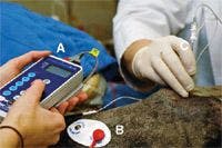

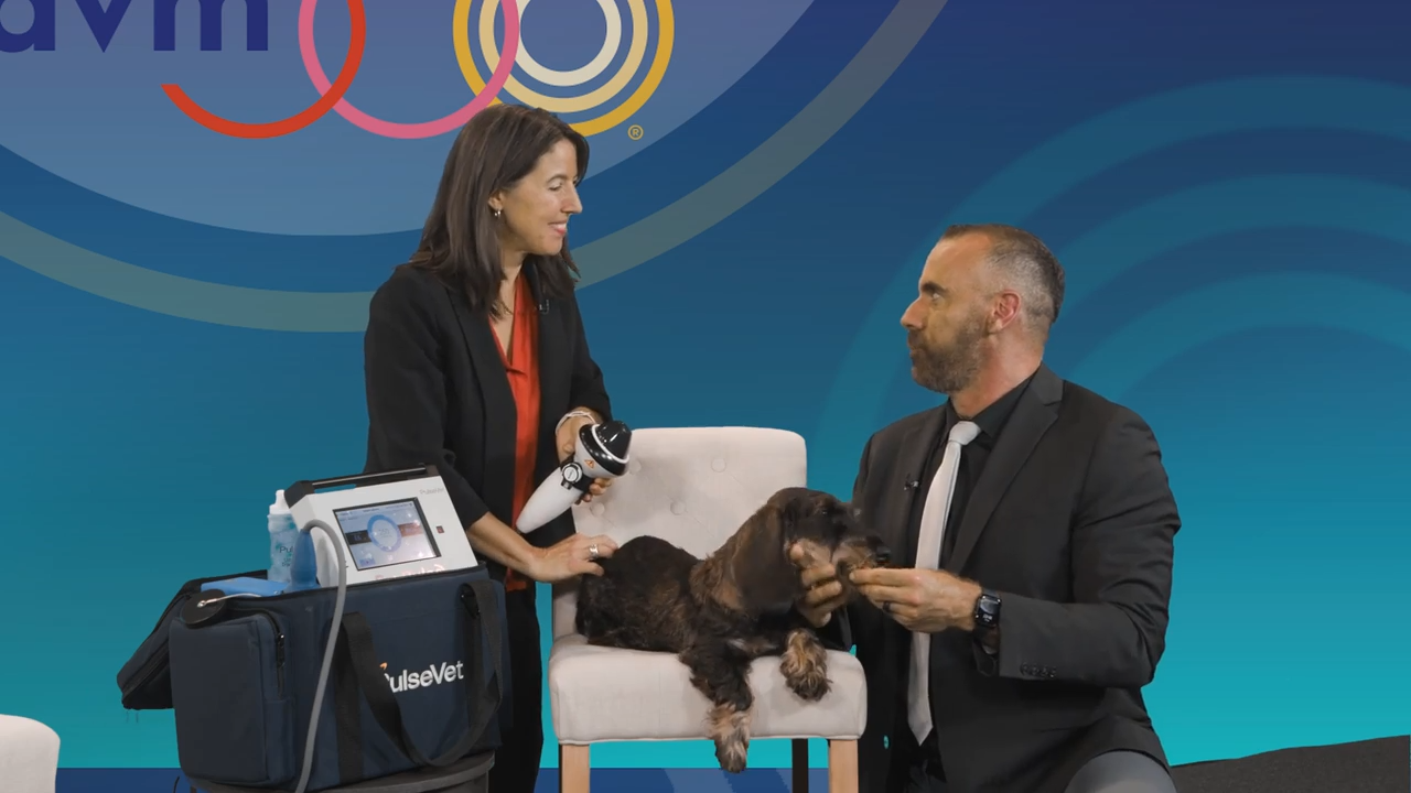
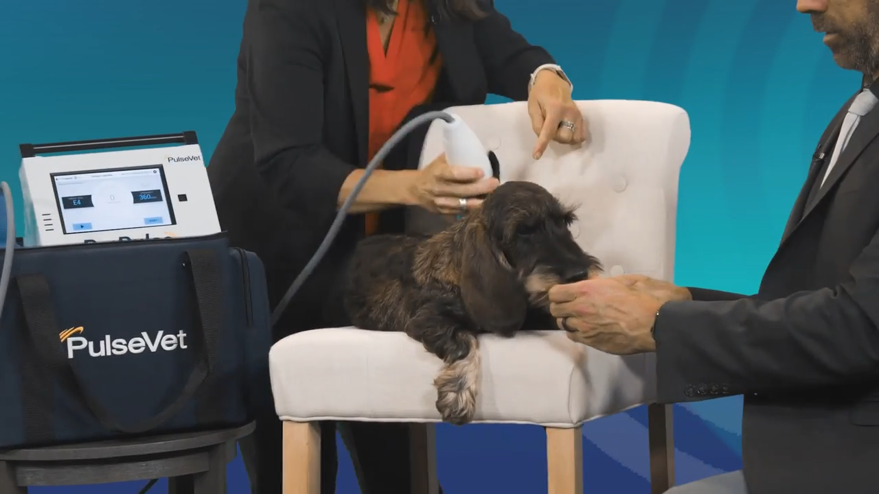
Figure 1 A. Using a nerve locator to block the sciatic nerve in a dog (A = current generator with control pad; B = grounding electrode; C = insulated stimulating needle).
Some controversy exists in the human literature regarding the appropriate amperage to optimize nerve proximity while avoiding intraneural or intrafascicular injection. Typically, the desired muscle response is present at 0.4 to 0.6 mA and absent at 0.2 mA.2,3 In addition, there should be no resistance to injection of the local anesthetic as this may indicate that the needle is within a nerve fascicle.3
Once proximity to a nerve is identified, a local anesthetic or mixture of local anesthetics and adjunctive agents can be injected and the needle can be removed. Alternatively, an indwelling perineural catheter can be placed for repeated or continuous administration of local anesthetics.
Christine Egger, DVM, MVSc, DACVA; Lydia Love, DVM, Department of Small Animal Clinical Sciences College of Veterinary Medicine The University of Tennessee Knoxville, TN 37996
REFERENCES
1. Mahler SP, Reece JLM. Electrical nerve stimulation to facilitate placement of an indwelling catheter for repeated brachial plexus block in a traumatized dog. Vet Anaesth Analg 2007;34(5):365-370.
2. Campoy L. Fundamentals of regional anesthesia using nerve stimulation in the dog. In: Gleed RD, Ludders JW, eds. Recent advances in veterinary anesthesia and analgesia: companion animals. Ithaca NY: International Veterinary Information Service, 2006 (www.ivis.org. Accessed Nov. 25, 2008.
3. Lemke KA, Crieghton CM. Paravertebral blockade of the brachial plexus in dogs. Vet Clin North Am Small Anim Pract 2008;38(6):1231–1241.