The pathophysiology of eosinophilic bronchopneumopathy
Eosinophilic bronchopneumopathy is thought to be a pulmonary hypersensitivity disorder that may be triggered by certain drugs, inhaled environmental allergens, or a parasitic, fungal, or bacterial respiratory tract infection.
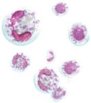
Eosinophilic bronchopneumopathy (EBP) is thought to be a pulmonary hypersensitivity disorder that may be triggered by certain drugs, inhaled environmental allergens, or a parasitic, fungal, or bacterial respiratory tract infection.1 However, the vast majority of cases are classified as idiopathic. The eosinophil is a primary player in the Type I hypersensitivity reaction that is speculated to generate this disease. Eosinophils compose about 2% of all circulating leukocytes and have a half-life in the tissues of about 12 days.2 Their principal function is to kill helminth parasites and tumor cells by releasing cytotoxins from intracellular granules.3


Another key player in allergic airway reactions in people is the T helper 2 (TH2) cell, and evidence indicates that this cell type may also predominate in the immune response that occurs in canine EBP.4 The TH2-type effector response is initiated when allergen is inhaled into the airways, promoting the expression of chemokines by lung epithelial cells.5 These chemokines recruit dendritic cells to the airways. Dendritic cells bind allergen on their surface via major histocompatibility complex II and then migrate to regional lymph nodes to present allergen to naïve CD4+ T cells.6 In fact, a predominance of CD4+ T cells has been documented in the bronchoalveolar lavage fluid of dogs with EBP.7 Interleukin-4 (IL-4), secreted by natural killer cells, basophils, and activated CD4+ T cells, induces differentiation of the naïve precursor T cells into TH2 cells in part by suppressing the TH1-type response.5
TH2 cells are characterized by their production of a specific cytokine profile including IL-4, IL-5, IL-9, and IL-13. IL-5 is essential for differentiation, activation, and survival of eosinophils, while IL-9 promotes mast cell accumulation within the airways.8,9 IL-4 and IL-13 have been shown to promote mucus hypersecretion, goblet cell hyperplasia, and subepithelial fibrosis in asthmatic disease.8 As part of the type I hypersensitivity reaction, IL-4 and IL-13 also induce B-lymphocytes to undergo isotype switching, resulting in the production of allergen-specific IgE.9 After allergen cross-links IgE on the surface of mast cells, these cells degranulate, releasing histamine, serotonin, and eicosanoids, as well as eosinophil chemotactic factors.3 Studies suggest that other chemotactic factors, such as eotaxin-2 and eotaxin-3 and monocyte chemoattractant protein-3, may play a significant role in recruiting eosinophils into the airways of dogs with EBP.10
The eosinophils that are attracted to the airways during an allergic response degranulate, releasing eosinophil cationic protein, eosinophil peroxidase, eosinophil-derived neurotoxin, and major basic protein.9 These cytotoxic proteins are specialized to destroy helminth parasites but probably mediate tissue damage within the airways when released in excess. In canine EBP, collagenases have been implicated as the enzymes that cause pulmonary tissue destruction.11 In particular, matrix metalloproteinase (MMP)-9 has been found in increased concentrations within the pulmonary macrophages and epithelial cells of dogs with EBP.12 Eosinophils may be involved in stimulating release of MMPs from these cells. Markers of collagenase activity that are increased in the bronchoalveolar lavage fluid of dogs with EBP include laminin-5 gamma 2-chain, a protein that is degraded by MMPs, resulting in epithelial cell sloughing, and procollagen type III amino terminal peptide, an amino acid sequence that is cleaved from the procollagen type III molecule during rapid collagen turnover.13,14
REFERENCES
1. Bauer T. Pulmonary hypersensitivity disorders. In: Kirk RW, ed. Current veterinary therapy X. Philadelphia, Pa.: WB Saunders, 1989;369-376.
2. Norris CR, Mellema MS. Eosinophilic pneumonia. In: King LG, ed. Textbook of respiratory disease in dogs and cats. Philadelphia, Pa.: WB Saunders, 2004;541-547.
3. Tizard IR. Type I hypersensitivity. Veterinary immunology: an introduction. 6th ed. Philadelphia, Pa.: WB Saunders, 2000;307-323.
4. Peeters D, Day MJ, Clercx C. Distribution of leucocyte subsets in bronchial mucosa from dogs with eosinophilic bronchopneumopathy. J Comp Pathol 2005;133(2-3):128-135.
5. Paul WE, Zhu J. How are TH2-type immune responses initiated and amplified? Nat Rev Immunol 2010;10(4):225-235.
6. Neurath MF, Finotto S, Glimcher LH. The role of Th1/Th2 polarization in mucosal immunity. Nat Med 2002;8(6):567-573.
7. Clercx C, Peeters D, German AJ, et al. An immunologic investigation of canine eosinophilic bronchopneumopathy. J Vet Intern Med 2002;16(3):229-237.
8. Finkelman FD, Hogan SP, Hershey GK, et al. Importance of cytokines in murine allergic airway disease and human asthma. J Immunol 2010;184(4):1663-1674.
9. Stone KD, Prussin C, Metcalfe DD. IgE, mast cells, basophils, and eosinophils. J Allergy Clin Immunol 2010;125(2 Suppl 2):S73-S80.
10. Peeters D, Peters IR, Clercx C, et al. Real-time RT-PCR quantification of mRNA encoding cytokines, CC chemokines and CCR3 in bronchial biopsies from dogs with eosinophilic bronchopneumopathy. Vet Immunol Immunopathol 2006;110(1-2):65-77.
11. Rajamäki MM, Järvinen AK, Sorsa T, et al. Collagenolytic activity in bronchoalveolar lavage fluid in canine pulmonary eosinophilia. J Vet Intern Med 2002;16(6):658-664.
12. Rajamäki MM, Järvinen AK, Sorsa T, et al. Clinical findings, bronchoalveolar lavage fluid cytology and matrix metalloproteinase-2 and -9 in canine pulmonary eosinophilia. Vet J 2002;163(2):168-181.
13. Rajamäki MM, Järvinen AK, Sorsa T, et al. Elevated levels of fragmented laminin-5 gamma2-chain in bronchoalveolar lavage fluid from dogs with pulmonary eosinophilia. Vet J 2006;171(3):562-565.
14. Schuller S, Valentin S, Remy B, et al. Analytical, physiologic, and clinical validation of a radioimmunoassay for measurement of procollagen type III amino terminal propetide in serum and bronchoalveolar lavage fluid obtained from dogs. Am J Vet Res 2006;67(5):749-755.