Paw pad pain: A review of corns in dogs
An overview of the clinical signs, potential causes, and treatment options available for corn-type lesions on canine paw pads.
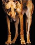
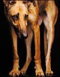
Sight hounds, especially greyhounds, often develop painful digital pad corn-type lesions. These well-demarcated, circumscribed hyperkeratotic lesions have a central core of keratin that is often conical. The lesions have the gross appearance of fibrous scar tissue and cause pain and local inflammation.1-4 They have been called corns,5-9 keratomas,3,4 and foot pad keratosis (orthokeratotic hyperkeratosis).10 The lesions are similar to the hard corn lesions seen on human feet (heloma durum), which are associated with chronic pressure or abrasion and overlie bony prominences where there is insufficient soft tissue between the skin and underlying bone.2,3

(GETTY IMAGES/ELKE VOGELSANG)
Corns can appear on the digital, metacarpal, or metatarsal paw pads, primarily in greyhounds, but most of the lesions occur on the digital paw pads. They are seen predominantly in active racing and retired middle-aged or older greyhounds.3,4,8-11
CLINICAL SIGNS
Clinical signs associated with corns include visible pain when palpation pressure is applied and a reluctance to walk on hard surfaces, both of which push the corn into subdermal tissues. It is important to apply pressure during palpation to elicit signs of pain.
There may be excessive nail growth on the affected paw, resulting from decreased wear as the dog tries to bear weight on the metacarpal or metatarsal pad rather than on the digital pads.3 When performing an orthopedic examination on a dog for lameness, especially a greyhound, a paw pad examination should be included.
ETIOPATHOGENESIS THEORIES
There are three theories as to the cause of corns:
1. Scar tissue and foreign bodies. One theory is that cuts or punctures of the pad result in an accumulation of scar tissue.6,7,9,12,13 The presence of a small foreign body in the pad is related to this theory. In this case, scar tissue accumulates at the lesion site as the body attempts to isolate the foreign body. This scar tissue develops into a thickened, hard corn on the pad.3,7,9,12,13
2. Papilloma virus infection. A second theory is that a corn lesion is the result of a papilloma virus infection.3,6,7,9 Because of the pressure and abrasion associated with ambulation, the papilloma does not grow on the pad surface. Instead, the lesion is forced into the deeper pad layers, resulting in a flat, circular, painful area (corn-type lesion) that is visible on the pad surface (Figure 1).6

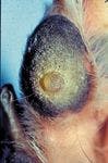
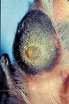
1. A corn lesion on a greyhound's digital pad.
3. Pressure and abrasion. A third theory for corn development is that repetitive mechanical trauma in the form of pressure and abrasion leads to lesion development (Wright JC, Borghese IF, Swaim SF, College of Veterinary Medicine, Auburn University, Ala: Unpublished data, 2003).3,7,9,10 These traumatic forces would be present in racing greyhounds. It has been reported11 and has been our observation that corns occur quite commonly in racing or retired greyhounds. However, lesions are also seen in whippets and lurchers.3
A look at the theories
The scar tissue, scar tissue and foreign body, and viral papilloma theories are probably not the cause of most corns in greyhounds for a variety of reasons.
Recurrence. Surgically removing the corn generally results in alleviating signs of pain; some dogs may be cured, and most remain corn-free for more than six months.3 Recurrence could be because of poor surgical technique, but it is more likely caused by underlying mechanical factors that have not been corrected (e.g. chronic low-grade pressure on the pad).3 The same reasoning holds true for the foreign-body-and-scar-tissue theory. If the foreign body has been removed, reaction or recurrence should not occur but it often does.
Lack of evidence. An electron microscopic examination of corns that had been removed did not show the presence of the papilloma virus.9 Likewise, immunohistochemical staining and polymerase chain reaction assay testing did not find evidence of viral etiology in corns removed from 18 greyhounds.1
Anatomic differences. Several suggestive factors support the pressure theory as a major cause of corns. One factor is the anatomic differences in the greyhound paw. Greyhounds have long, narrow paws with little distance between pads, which results in greyhounds experiencing more ground reaction forces than dogs with wider paws.14 The average velocity of a medium-grade racing greyhound is 36 miles per hour (extrapolated from track data), and photographic evidence shows that during a gallop, the dog is airborne and lands initially on the extended paw of a thoracic limb on a firm sand surface. As a result, ground reaction force is high.3
Sparsity of tissue. It has also been theorized that since greyhounds have a sparsity of subcutaneous adipose tissue throughout their bodies, a sparsity of fibroadipose padding tissue may also exist in the paw pads. Thus, the distal interphalangeal articulation of the digit is in closer apposition to the dermis of the pad skin. And, thus, when the dog ambulates, pressure on the pad skin by this joint causes corn-type lesions to form.3,7,9,10
Anatomic abnormalities. The mechanical theory is supported by the presence of anatomical abnormalities, resulting in abnormal weight bearing on the pad. Deep digital flexor tendon rupture or stretching is a common finding. Also if the anatomic abnormality can be corrected then the corn will disappear.3
Weight-bearing digits primarily affected. Another factor that supports the pressure theory is that digits 3 and 4, the major weight-bearing digits, are primarily involved. In a study of 30 dogs with a total of 40 corns, 36 of the corns were on digits 3 and 4 of the thoracic limbs.3 In another study of 24 dogs—18 of which were greyhounds—24 of 26 corns were on digits 3 and 4.1
In a national epidemiologic survey of 484 greyhound owners, 275 male greyhounds and 209 female greyhounds were evaluated for corns (Wright JC, Borghese IF, Swaim SF, College of Veterinary Medicine, Auburn University, Ala: Unpublished data, 2003). The median weight of the dogs was 70 lb (31.8 kg) with a range of 44 to 104 lb (20 to 47.3 kg). One hundred seventy-five owners reported that their dogs had corn-type lesions, and 121 of the owners indicated the location of the corns on the digits. The survey asked the greyhound owners to report any racing injuries, if known. The responses indicated that no significant racing injuries were associated with the occurrence of corns.
The corn lesions were not associated with either the sex or the weight of the dogs, and there was no significant difference in location among paws (Wright JC, Borghese IF, Swaim SF, College of Veterinary Medicine, Auburn University, Ala: Unpublished data, 2003). However, digits 3 and 4 had a significantly higher occurrence of lesions (Table 1). Thus, from a wide geographic area and a large population of greyhounds, the same phenomenon was evident—predominance of corns on the primary weight-bearing digits.

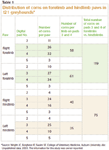

Table 1: Distribution of corns on forelimb and hindlimb paws in 121 greyhounds*
There appears to be a predilection for corns to occur on the paws of the thoracic limbs. This tendency was noticed in the epidemiologic survey of greyhound owners (Table 1) and may be related to ground reaction forces.3 In dogs, the vertical forces placed on the thoracic limb paw pads during walking are about 1.1 times body weight; whereas, on the pelvic limb pads, this force is 0.8 times body weight.15
TREATMENT OPTIONS AND PREVENTION
One author (Dr. Swaim) has noticed that the number of treatments that have been described for a condition is an indication of the problematic nature of the condition. Digital pad corns are an example of this phenomenon. Numerous treatments for digital pad corns have been described (see "Potential treatment options for digital pad corns"). However, few treatments for digital pad corns have been described in scientific literature.


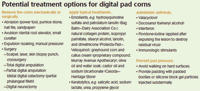
Potential treatment options for digital pad corns
Two of the authors (Dr. Swaim, Dr. Bohling) use surgical excision of the corn as the initial part of treatment. A No. 15 scalpel blade is used to make an elliptical incision around the corn. The corn is removed with blunt and sharp dissection. Since corns are composed of dense scar-type tissue and confined to the dermis of the pad skin, their edges are well-defined to make removal easier (Figure 2). Digital pressure is used to attain hemostasis. Closure of the resulting defect is with interrupted far-near-near-far, 3-0 monofilament nylon or polypropylene sutures.

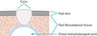
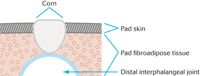
2. Diagrammatic representation of a cross section of a corn.
A pressure-relief bandage is then placed on the paw. To make a pressure-relief bandage, the paw is first wrapped in a routine paw bandage. Next, a triangular piece of foam sponge (Confor Foam—Hi-Tech Foams) the size of the metacarpal or metatarsal pad is placed under the bandaged paw. The cup portion of an appropriate-sized metal Mason metasplint is then placed over the foam sponge, holding it to the bandage. Then this is bandaged in place. This arrangement elevates the digital pads to relieve pressure on them as the incision heals.16 The bandage is changed periodically during a 14-day healing period. Sutures are removed 14 days after surgery.
Pressure prevention can be approached by advising owners to keep their dogs from walking on hard surfaces and to place padded booties on their dogs' paws.
FUTURE TREATMENT OPTIONS
Two approaches could be considered for relieving local pressure: providing local padding and removing pressure points.
For people, local padding comes in the form of corn pads, which are donut-shaped foam rubber pads with an adhesive surface placed with the hole over the offending prominence. For dogs, however, rarely would such pads be tolerated, and their durability would not be sufficient.
In people, injecting liquid silicone under pressure points in their feet has resulted in plantar pressure relief.17,18 A modification of this technique has been studied preclinically in dogs. Silicone block gel particles were implanted subdermally under the digital pads. Ground contact pressure analysis revealed a decrease in pressure three months after implantation; however, there was some migration of the particles.9
In a clinical study conducted by one of the authors (Dr. Bohling; unpublished data), five affected greyhounds received silicone block gel particle implants under the affected digital pads. All five dogs had complete resolution of signs of pain and lameness. However, with weight bearing, the implants eventually migrated away from the area of greatest pressure, and corns recurred in all dogs within about six months after implantation.
This result led to work to develop an implant in which the silicone block gel particles are encapsulated. Preliminary clinical trials of this implant have been performed in five dogs (Dr. Bohling; unpublished data). One dog rejected the implants, and in one dog, the particles migrated because of a damaged implant. Three dogs experienced no complications and were still clinically normal (pain-free with no lameness) at last contact, up to two years after implantation.
To pursue the theory that pressure from the distal interphalgeal joint on the pad skin is causing the problem, research could be performed to further develop the silicone block gel particle encapsulation technique and to develop an arthroplasty technique, in which the joint is removed and replaced with a cushioning material (e.g. medical grade silicone).
SUMMARY
When examining dogs, especially greyhounds, for lameness, be sure to include paw pad examinations. Finding a corn that is painful on pressure application is important and must be addressed. Two reports in the literature1,3 and the study done at Auburn University's College of Veterinary Medicine (Wright JC, Borghese IF, Swaim SF, College of Veterinary Medicine, Auburn University, Ala: Unpublished data, 2003) indicate that removing the corn and preventing digital pad pressure during healing is the most efficacious treatment.1,3 Further research is needed in perfecting and developing local pressure prevention techniques.
Editors' note: Ilaria F. Borghese is the president of Thera-Paw, Inc., and CEO of Symposium on Therapeutic Advances in Animal Rehabilitation.
Steven F. Swaim, DVM, MS
Scott-Ritchey Research Center and Department of Clinical Sciences
College of Veterinary Medicine
Auburn University
Auburn, AL 36849
Mark W. Bohling, DVM, PhD, DACVS
Regional Institute for Veterinary Emergencies and Referrals
2132 Amnicola Highway
Chattanooga, TN 37406
James C. Wright, DVM, PhD, DACVPM
Department of Pathobiology
College of Veterinary Medicine
Auburn University
Auburn, AL 36849
Ilaria F. Borghese, MS, MA, OTR/L
Thera-Paw, Inc.
36 Hill and Dale Road
Lebanon, NJ 08833
To view the references for this article, visit dvm360.com/CornRefs.