Pneumopericardium associated with peritoneopericardial diaphragmatic hernia repair in a dog
A hernia repair leads to this unusual radiographic finding.
A 22-month-old intact male Shih Tzu was referred for further investigation and treatment of intermittent vomiting and exercise intolerance of four weeks' duration. A thoracic radiographic examination showed cardiomegaly and intestinal loops in the pericardial sac (Figure 1).

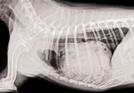

Figure 1. A lateral thoracic radiograph of a peritoneopericardial diaphragmatic hernia in a 22-month-old Shih Tzu.A peritoneopericardial diaphragmatic hernia (PPDH) was diagnosed, and the dog underwent surgical repair through a ventral midline celiotomy. The dog recovered successfully from the surgery.
DIAGNOSTIC IMAGING
Postoperative thoracic radiographs were obtained four hours after surgery to check the position of the central venous pressure catheter placed in the left jugular vein (Figures 2A & 2B). The radiographic examination revealed a well-demarcated region of gas opacity surrounding the cardiac silhouette that was devoid of pulmonary vascular or airway markings. This radiolucent area was well-delineated by an opaque line representing the pericardium. The apex of the cardiac silhouette was displaced from the sternum.

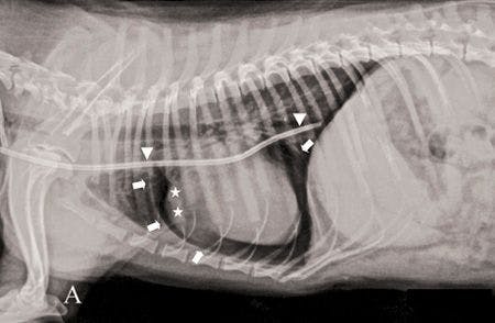


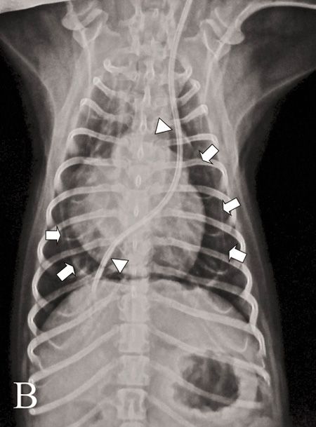
Figures 2A &2B. Right lateral (A) and dorsoventral (B) thoracic radiographs obtained four hours after PPDH repair. Note the pericardium (white arrows), a pneumopericardium (radiolucent area inside the pericardium), a prominent right atrium and distended right auricle (asterisks), and a catheter placed in the cranial and caudal vena cava through the left jugular vein for central venous pressure measurement (arrowheads).A prominent right atrium and distended right auricle were clearly defined. A catheter placed in the cranial and caudal vena cava through the left jugular vein for central venous pressure measurement was also observed. Pneumopericardium was diagnosed. Pneumoperitoneum and loss of abdominal serosal detail were also evident.
TREATMENT AND FOLLOW-UP
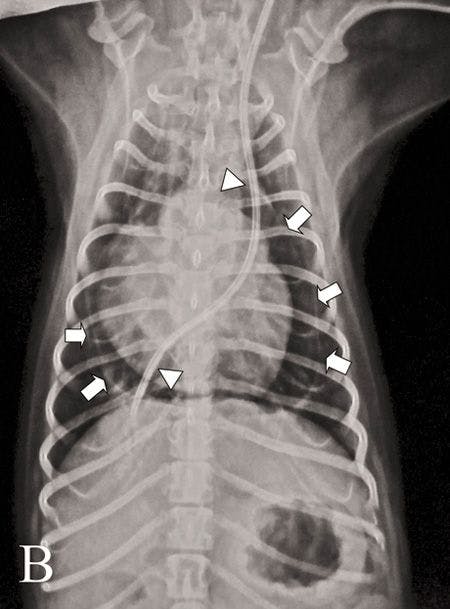
No specific treatment was required. Recheck radiographic examination of the thorax was performed five (Figure 3) and 30 days after surgery, which confirmed resolution of pneumopericardium. No further abnormalities were detected. Eleven months after surgery, the dog is doing well.

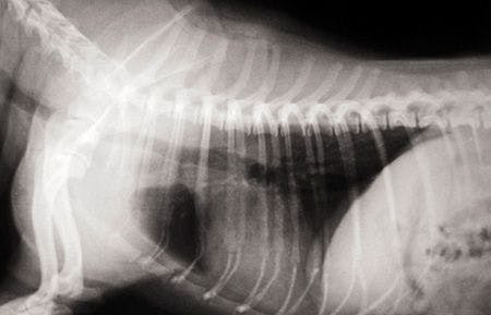
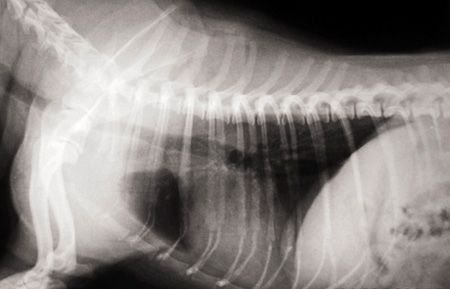
Figure 3. A lateral thoracic radiograph obtained five days after surgery for a PPDH repair. The pneumopericardium had resolved.DISCUSSION
Pneumopericardium, by definition, is an accumulation of free gas within the pericardial sac. This condition is not commonly reported in dogs and cats.1-6 Pneumopericardium may be caused by trauma or may be nontraumatic. Reported causes in small animals include thoracic trauma, positive pressure ventilation, pulmonary-pericardial communication, tracheal rupture related to intubation, and alveolar rupture associated with cough and bronchospasm.1-6
Clinical signs of pneumopericardium may depend on the volume and rate of air introduced into the pericardial sac. Pneumopericardium associated with PPDH repair has not been reported in the veterinary literature.
PPDH
PPDH is a congenital anomaly in dogs that allows communication between the pericardial sac and the peritoneal cavity through a diaphragmatic defect.7-9 Abdominal viscera that can be herniated into the pericardial sac include liver, gallbladder, omentum, intestines, spleen, and pancreas.7,9 Adhesions between the herniated organs and pericardium or myocardium may also be seen.7 Many dogs do not exhibit clinical signs, and PPDH is an incidental finding during routine physical or radiographic examination.7,9
Common clinical signs include vomiting, respiratory distress, weight loss, anorexia, lethargy, exercise intolerance, collapse, fever, and diarrhea.7,9 Physical examination findings may be normal or may detect muffled heart sounds, thoracic borborygmi, decreased lung sounds, tachypnea, or signs secondary to cardiac tamponade.7-9 PPDH is diagnosed by plain radiography; barium administration, ultrasonography, echocardiography, and computed tomography may also be used to confirm diagnosis. In the present case diagnosis was made by plain radiography, and no barium study was performed.
Surgical treatment is recommended in dogs with clinical signs.7-9 Closure of the diaphragmatic defect with sutures is performed through a ventral midline celiotomy. Postoperative complications are considered minor in most dogs after PPDH repair.7
Pneumopericardium
Pneumopericardium might be a potential complication of PPDH repair.8 The volume of air accumulated within a very big pericardial sac may depend on the size of the diaphragmatic defect; resultant pneumopericardium may interfere with cardiopulmonary function and should be evacuated by centesis through the diaphragm soon after diaphragmatic closure.8,10
In the present report, pneumopericardium was created because the remaining air in the pericardial sac was not removed by centesis after diaphragmatic closure. Experimental progressive pneumopericardium in dogs was reported to result in cardiac tamponade, producing serious hemodynamic changes similar to those related to pericardial effusion.10 Our dog, however, recovered uneventfully from surgery. After 48 hours of hemodynamic monitoring, the dog did not show any clinical signs related to pneumopericardium, which was diagnosed incidentally during routine imaging.
Most of the pneumopericardium clinical cases reported required no specific treatment, and all resolved spontaneously.1,2,4-6 In one case, however, lung lobectomy for the correction of a pulmonary-pericardial communication was performed in a dog that was exhibiting no clinical signs but had a pneumopericardium to prevent the potential development of cardiac tamponade.3
In the present report, pneumopericardium and lack of pericardial constraint allowed the right atrium and auricle to expand beyond their normal volume. Other congenital deformities may accompany PPDH.9 In this case, the central venous catheter that was placed through the left jugular vein entered the cranial vena cava, which is on the left side of the heart instead of the normal right side, suggesting a possible persistent left cranial vena cava.11 Persistent left cranial vena cava is uncommonly reported in dogs, coexisting or not with other cardiac defects.11
Echocardiography and nonselective angiography should be performed to make the diagnosis and exclude other cardiac defects.11 The presence of a persistent left cranial vena cava may lead to confusion if thoracic surgery or other angiographic procedures are going to be performed. No further imaging, however, was performed to confirm this diagnosis in the present report.
Conclusion
Pneumopericardium may be an interesting phenomenon that most surgeons might not be aware of. In this case, it was only detected because we chose to take four-hour postoperative radiographs for other reasons, something most surgeons would not consider clinically indicated. In our case, the pneumopericardium was a radiographic finding of minor clinical significance, which did not require treatment as it did not impede cardiopulmonary function.
References
1. Brown DC, Holt D. Subcutaneous emphysema, pneumothorax, pneumomediastinum, and pneumopericardium associated with positive-pressure ventilation in a cat. J Am Vet Med Assoc 1995;206(7):997-999.
2. Parent C, Rozanski E. What is your diagnosis? Pneumopericardium and pulmonary alveolar disease (consistent with pulmonary contusions). J Am Vet Med Assoc 1998;212(9):1377-1378.
3. Leclerc A, Brisson BA, Dobson H. Pneumopericardium associated with a pulmonary-pericardial communication in a dog. J Am Vet Med Assoc 2004;224(5):710-712.
4. Johnson-Neitman JL, Huber ML, Amann JF. What is your diagnosis? Pneumomediastinum and pneumopericardium. J Am Vet Med Assoc 2006;229(3):359-360.
5. Orlando JM. What is your diagnosis? Pneumopericardium and bilateral pneumothorax in a Labrador Retriever. J Am Vet Med Assoc 2009;235(10):1145-1146.
6. Agut A, Costa-Teixeira MA, Cardoso L, et al. What is your diagnosis? Pneumopericardium. J Am Vet Med Assoc 2010;237(4):363-364.
7. Banz AC, Gottfried SD. Peritoneopericardial diaphragmatic hernia: a retrospective study of 31 cats and eight dogs. J Am Anim Hosp Assoc 2010;46(6):390-404.
8. Hunt GB, Johnson KA. Diaphragmatic hernias. In: Tobias KM, Johnston SA, eds. Veterinary surgery small animal. St. Louis: Elsevier, 2012;1380-1390.
9. Wallace J, Mullen HS, Lesser MB. A technique for surgical correction of peritoneal pericardial diaphragmatic hernia in dogs and cats. J Am Anim Hosp Assoc 1992;28(2):503-510.
10. Reed JR, Thomas WP. Hemodynamics of progressive pneumopericardium in the dog. Am J Vet Res 1984;45(2):301-307.
11. Fernandez del Palacio MJ, Bayon A, Agut A. Dilated coronary sinus in a dog with persistent left cranial vena cava. Vet Radiol Ultrasound 1997;38(5):376-379.
Lysimachos G. Papazoglou, DVM, PhD, MRCVS
Michail N. Patsikas, DVM, MD, PhD, DECVDI'
Anna Deligianni, DVM
Georgios Kazakos, DVM, PhD
Department of Clinical Sciences
Faculty of Veterinary Medicine
Aristotle University of Thessaloniki
Thessaloniki, Greece
Erik R. Wisner, DVM, DACVR
Department of Surgical and Radiological Sciences
School of Veterinary Medicine
University of California
Davis, CA 95616