Practical Matters: Use comparison radiographs when identifying orthopedic lesions in young dogs
A useful protocol for practitioners who review their own films or send them to a specialist is to routinely obtain orthogonal views of the affected and normal bones.
Traumatic injuries in young dogs can produce subtle radiographic changes that are confusing and difficult to identify because of open physes and whole body (vs. collimated or focused) radiographic examinations. Radiographic characteristics such as a displaced bone fragment, a narrowed or widened physis, and periosseous soft tissue thickening can be hard to recognize. A useful protocol for practitioners who review their own films or send them to a specialist is to routinely obtain orthogonal views of the affected and normal bones. In many patients, side-by-side imaging using the same view of each bone will further help veterinarians identify a lesion (Figures 1 & 2). Because veterinary patients tend to be quite mobile, sedation is often necessary for accurate imaging.

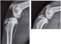

Figure 1 & 2. Lateral radiographs of the right (1) and left (2) stifle joints in a 4-month-old keeshond. The radiograph of the right stifle reveals no abnormalities; the radiograph of the left stifle reveals an avulsion fracture of the tibial tuberosity. (Case courtesy of Dr. Kevin French.)
In addition, common injuries, such as avulsion of the tibial tuberosity apophysis, a proximal femoral capital physeal fracture, and a distal humeral or femoral condyle fracture, can be more easily appreciated by clients when a radiograph of the injury is compared with a radiograph of the normal bone. Furthermore, with an injury of several weeks' duration in immature dogs, radiographic examination of the contralateral limb is critical in evaluating any disturbance of longitudinal bone growth.


Joseph Harari, MS, DVM, DACVS
Joseph Harari, MS, DVM, DACVS
Veterinary Surgical Specialists
21 E. Mission Ave.
Spokane, WA 99202


 in general practice")


