The role of topical therapy in the successful treatment of allergic dermatitis
Topical therapy is an important - and in many cases essential - component of successfully managing allergic dermatitis in dogs. When used as an adjunctive treatment for generalized disease, topical therapy often minimizes dependence on systemic medications that may be deleterious to the patient's health. In addition, topical therapy may be more effective in treating localized or regionalized pruritus.
Topical therapy is an important—and in many cases essential—component of successfully managing allergic dermatitis in dogs. When used as an adjunctive treatment for generalized disease, topical therapy often minimizes dependence on systemic medications that may be deleterious to the patient's health. In addition, topical therapy may be more effective in treating localized or regionalized pruritus.
The development of new products with improved active ingredients and delivery systems has made topical therapy even more effective. The same active ingredients are often used in different delivery systems, such as shampoos, rinses, powders, lotions, sprays, creams, emulsions, ointments, and gels. The advantages and disadvantages of these formulations vary, as do their cost, ease of application, and efficacy.
Shampoos and leave-on products
Shampoo therapy is the cornerstone of the successful topical management of an allergic patient. Frequent bathing with the correct product helps control pruritus by removing offending allergens and desensitizing and moisturizing the skin. Critical factors affecting the efficacy of shampoo therapy include client compliance, contact time, frequency of use, and active ingredients.
Client compliance is the most important component of successful shampoo therapy. Practitioners should train themselves and their technicians to educate clients about contact time and frequency of use to increase the success rate for allergic patients. Practitioners must stress that effective shampoo therapy requires a minimum of 10 contact minutes before rinsing to allow sufficient product interaction with the skin. Remember that the longest 10 minutes of a dog owner's life are the 10 minutes spent lathering his dog in a bathtub!
It's also important to discuss frequency of shampoo therapy with owners. Although frequency generally depends on the specific condition, shampooing once or twice weekly is usually necessary to successfully manage a pruritic patient. When practitioners educate clients about why frequency of use and contact time are essential, compliance increases.


Table 1. Topical Antipruritic Ingredients and Products
In addition to shampoos, products that are left on the skin, such as lotions, creams, and sprays, may provide added advantages. These products can often be used in conjunction with appropriate shampoo therapy. Most of the active ingredients in shampoos are also available in these leave-on formulations. The main disadvantage of using these products is that they require direct skin contact. Therefore, they are more beneficial in short-coated dogs or in areas where the hair can be clipped or parted easily, such as the paws, axillae, inguina, and ventral abdomen.
Active ingredients
Practitioners should familiarize themselves with several important ingredients of antipruritic products (see Table 1 for products containing these ingredients):
Colloidal oatmeal, fatty acids, urea, glycerin, and other moisturizers help alleviate skin dryness through humectant properties. This subsequently allows for an increase in the pruritic threshold of the skin.
Menthol, camphor, and thymol act by substituting other sensations for the pruritus. A cooling sensation helps decrease pruritus and may also help increase the pruritic threshold.
Pramoxine, a local anesthetic for skin nerve endings, may also have antipruritic properties that have not yet been completely determined.1 Studies have shown that pramoxine is safer than other topical anesthetics.1
Lidocaineis another local anesthetic that affects peripheral nerves and can be used safely in topical products.
Diphenhydramine, an antihistamine, can have mild antipruritic effects when administered topically. It has been shown to cross the epidermal barrier to exert its antihistaminic effects.1
Hydrocortisone and other more potent long-acting cortisones, such as betamethasone, can have anti-inflammatory and antipruritic properties. Hydrocortisone is the safest glucocorticoid for topical use but is also the least potent when applied topically. On the other hand, the fluorinated cortisones, including betamethasone, are the most potent but have the greatest potential for systemic side effects. Several studies have demonstrated that corticosteroids applied topically to the skin and eye can suppress the adrenal axis and elevate hepatic enzyme activity.1 Therefore, topical glucocorticoids should not be considered innocuous, and practitioners should approach long-term application with care and closely monitor the results.
Triamcinolone acetonide (0.015%) is the active ingredient in Genesis Topical Spray (Virbac), which also can be useful in clinical settings. The spray is available in 8- and 16-oz sizes. In this formulation, triamcinolone is a more potent corticosteroid than hydrocortisone, and I have found it to be more effective for allergy-induced pruritus. Furthermore, it is less potent than products containing betamethasone, fluocinolone, and dexamethasone; therefore, it may cause fewer side effects. Studies have demonstrated the efficacy of triamcinolone topical spray in both experimental models and in a controlled, double-blind placebo study conducted in a clinical setting.2,3 In the latter study, pruritus in 67% of allergic dogs (35 of 52) was successfully controlled as defined by the study parameters.3 Treated dogs exhibited no significant abnormalities on serum chemistry panels or complete blood counts and fewer adverse clinical effects than dogs treated with placebo.3
Clinical applications
The key to a successful topical therapy regimen is appropriate case selection. Knowing when to choose specific products for maximum efficacy takes experience, logic, and thought. Pruritus is a clinical sign of many diseases, and not all pruritic patients suffer from allergies as a component of their dermatologic disease. Picking antipruritic products while ignoring the entire clinical picture will lead to decreased effectiveness of the product for the patient and frustration on the part of the practitioner and client.
To look at the whole clinical picture, practitioners must:
- recognize and manage secondary problems, if present, such as bacterial pyoderma and Malassezia dermatitis with appropriate antibiotic and antifungal therapy
- eliminate the possibility of other diseases, such as demodicosis, dermatophytosis, scabies, or cheyletiellosis
- address the possibility of additional allergies, such as contact, flea bite, or food allergies.
Therefore, practitioners should take a thorough history, perform a complete physical examination, and undertake routine diagnostics, such as skin scrapings, cytologic impression smears, and dermatophyte cultures, for every patient that presents with pruritus as a clinical sign. Aggressive flea control is warranted, and in cases of nonseasonal pruritus, prophylactic therapy against parasitic mites should be considered. In addition, practitioners should perform appropriate elimination food trials in cases of nonseasonal pruritus to rule out food allergy dermatitis. Remember, launching blindly into therapy without recognizing all other aspects of the patient's condition usually results in short-term control at best or outright failure.
If pruritus persists after secondary factors have been appropriately managed and other diseases have been eliminated from the differential list, topical antipruritic therapy may be indicated to manage the residual allergic pruritus. While allergic patients of all types can benefit from appropriate antipruritic topical therapy, it is generally more effective in managing atopic patients.
Atopy is a heritable inflammatory and pruritic allergic skin disease with characteristic clinical features. It is most commonly associated with IgE antibody production in response to environmental allergens.1 The route of exposure to these environmental allergens is thought to be both from inhalation and percutaneous absorption.4 The typical age of onset is between 6 months and 3 years.5 Many breeds have an increased incidence, but this may reflect regional biases and breed popularities. Pruritus can be seasonal or nonseasonal, depending on which allergens the patient is allergic to; however, seasonality has been reported to initially occur in up to 75% of dogs.5 The most common areas affected include the face, ears, paws, axillae, legs, and ventral abdomen, although generalized pruritus may also be seen. It is also important to recognize that very few primary lesions are present in pure atopic cases; only secondary lesions are commonly seen and may include erythema, scales, and excoriations. If primary lesions such as papules, pustules, or vesicles are noted diffusely, additional diagnostics and therapy as discussed earlier are warranted before practitioners can tentatively diagnose atopy.

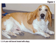

Figure 1. A 4-year-old basset hound with atopy.
Once the clinician confidently diagnoses atopic dermatitis and manages the secondary problems, topical therapy may effectively control pruritus. The following case study describes one way to manage an atopic dog using topical therapy.
Case study
A 4-year-old basset hound presented with a two-year history of nonseasonal pruritus involving the paws, legs, axillae, and ventral abdomen (Figure 1). Recurrent ear infections had been treated in the past. No other dogs in the house were pruritic, and the owner was unaffected. The owner used monthly flea control on the dog and had an exterminator treat the house and environment every six months. A previous strict food trial with a duck and potato diet was used for one year with no reduction in pruritus. There had been no response to various antihistamines, and although injectable corticosteroids had been effective, they had not been used in the last six months.

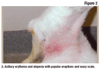

Figure 2. Axillary erythema and alopecia with papular eruptions and waxy scale.
Dermatologic examination revealed generalized papular to crusted pustular eruptions across the trunk with patchy alopecia in the affected areas (Figures 2 and 3). All four paws were erythematous interdigitally. The rest of the physical examination was within normal limits.
Cytologic impression smears of the papules on the trunk and the interdigital areas of the paws revealed cocci (likely Staphylococcus intermedius) extracellularly and intracellularly within neutrophils. Skin scrapings were negative for parasites, and dermatophyte cultures were taken from the papular eruptions. To control the bacterial population, oral cephalexin was prescribed at a dosage of 22 mg/kg twice daily for three weeks. Twice weekly bathing with an antibacterial shampoo was prescribed as an adjunctive therapy to help resolve the pyoderma.
Upon reexamination three weeks later, the dog exhibited a 50% reduction in pruritus. The remaining pruritus was noted on the paws, axillae, and ventral abdomen. Dermatophyte cultures were negative. The papular and pustular eruptions had resolved, and the dog was sedated for intradermal skin testing. Numerous positive reactions were noted, and immunotherapy was formulated and instituted based on the results. However, given that immunotherapy may take several months to become effective and reduce clinical signs, a topical approach was instituted to help control the dog's remaining pruritus.
Figure 3. Ventral abdominal and inguinal erythema and mild alopecia with papular eruptions.
My recommendations for home care included twice weekly bathing using oatmeal-based shampoo with 10 minutes of skin contact time followed by moisturizing rinses to help hydrate the skin. In addition, triamcinolone topical spray (twice daily for seven days then on an as-needed basis) was used on the paws and ventral abdomen. With this topical therapy protocol, the dog was managed comfortably until immunotherapy became effective over the next few months. (Note: Although Genesis is labeled for use for only 28 days, many dermatologists use it for longer periods in some cases, as we did with this dog. With proper patient monitoring and appropriately informed clients, extralabel use of topical triamcinolone can be beneficial in many patients.)
Owners can easily apply triamcinolone spray at home, although they should wear gloves. For best results, the product should be applied twice daily for the first week, once daily for the second week, and on alternating days thereafter. It is labeled for use for up to 28 days. Product application should be demonstrated to the client by a trained technician to illustrate the importance of achieving thorough skin contact and to improve client compliance. The hair should be parted and the area sprayed until the affected skin is uniformly wet without any runoff. The solution should then be massaged into the skin with a gloved hand.
Conclusion
Topical therapy is a safe and effective tool in managing chronic pruritus associated with allergic dermatitis. Client compliance is the most important component of a successful topical therapy protocol. Adequate client education helps ensure compliance and successful treatment. There are a number of topical therapy products containing various active ingredients. Therefore, practitioners must pay special attention to the specific product to make the best choice for the individual patient.
References
1. Muller GH, Kirk RW, Scott DW. Small Animal Dermatology. 6th ed. Philadelphia, Pa: WB Saunders Co, 2001;228-237.
2. DeBoer DJ, Cooley JA. Use of induced cutaneous immediate-type hypersensitivity reactions to evaluate anti-inflammatory effects of triamcinolone topical solution in three dogs. Vet Dermatol 2000;11:25-37.
3. DeBoer DJ, Schafer JH, Salsbury CS, et al. Multiple-center study of reduced-concentration triamcinolone topical solution for the treatment of dogs with known or suspected allergic pruritus. Am J Vet Res 2002;63:408-413.
4. Olivry T, Hill PB. The ACVD task force on canine atopic dermatitis (IX): The controversy surrounding the route of allergen challenge in canine atopic dermatitis. Vet Immunol Immunopathol 2001;81:219-225.
5. Griffin CE, DeBoer DJ. The ACVD task force on canine atopic dermatitis (XIV): Clinical manifestations of canine atopic dermatitis. Vet Immunol Immunopathol 2001;81:255-269.