Salivary mucoceles in cats: A retrospective study of seven cases
Salivary mucoceles are formed by the extravasation and accumulation of saliva in tissues adjacent to a salivary gland. The accumulated saliva incites an inflammatory response, leading to a walled-off accumulation of mucoid fluid.
Salivary mucoceles are formed by the extravasation and accumulation of saliva in tissues adjacent to a salivary gland. The accumulated saliva incites an inflammatory response, leading to a walled-off accumulation of mucoid fluid. Trauma can cause salivary mucoceles; however, most cases are idiopathic.1
Salivary mucoceles are uncommon in cats. Although individual case reports of feline salivary mucoceles exist,2-8 no case series has been reported in the veterinary literature.
In this article, we present findings from our review of the records of seven cats with salivary mucoceles to help identify common presenting signs and the best form of treatment.
SALIVARY MUCOCELES: AN OVERVIEW
In cats, salivary mucoceles range from nonclinical to life-threatening in severity.2-8 Cats have five major salivary glands (parotid, mandibular, sublingual, molar, zygomatic) compared with dogs' four (parotid, mandibular, sublingual, zygomatic),9,10,11 and mucoceles in cats have been associated with all but the molar gland.2-8 In dogs, the most commonly affected gland is the sublingual gland.12 However, the small number of cases in cats makes determining the most commonly affected salivary gland difficult.


PREVIEW
Clinical signs and diagnosis
Clinical signs of mucoceles include a fluctuant, nonpainful mass close to the salivary glands, dysphagia, exophthalmia, and dyspnea. Signs largely depend on the location and size of the mucocele. Differential diagnoses include an abscess, neoplasia, a foreign body, and a granuloma.
Diagnosis involves aspiration of a thick, stringy, golden fluid with low cellularity from the mass.1,13 Sialography has been performed in dogs and may be useful for identifying specific gland involvement when it is not readily apparent.12,13 Other diagnostic imaging tests (radiography, ultrasonography, computed tomography) may help rule out other disease processes, such as a foreign object or involvement or invasion of multiple tissues, but are unlikely to definitively identify a salivary mucocele.14,15
Treatment
Treatment options include draining the mucocele, marsupialization, and removing the involved gland. Drainage alone is likely to result in recurrence unless the mucus source is addressed. Marsupialization, which involves excising an elliptical section of mucosa over the mucocele, draining the saliva, and apposing the mucosal surface to underlying connective tissue, does not address the underlying problem. And because of potential complications such as frequent recurrence and the fact that future surgical intervention would be difficult, it is not the preferred procedure.1,13,16 Thus, surgically excising the affected gland is ideal.
CRITERIA FOR CASE SELECTION
The medical records of all cats examined for further management of a salivary mucocele at the University of Pennsylvania Matthew J. Ryan Veterinary Hospital and Red Bank Veterinary Hospital between 1993 and 2006 were reviewed. Cats with confirmed salivary mucoceles that were surgically repaired were included in this case series. Seven cats met these criteria.
PROCEDURES AND RESULTS
Each cat's history; signalment; physical examination, complete blood count (cbc), and serum chemistry profile results (when available); clinical signs; treatment; and outcome were identified and recorded. The signalment and location and surgical correction of the mucoceles for the seven cats are shown in Table 1.


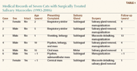
Table 1: Medical Records of Seven Cats with Surgically Treated Salivary Mucoceles (1993-2006)
History
Three of the cats were periodically allowed outdoors, and four were strictly indoor cats. Five of the seven cats had been seen by a referring veterinarian and treated with antibiotics (ampicillin, amoxicillin trihydrate-clavulanate potassium) with no improvement of clinical signs.
Physical examination findings
The mass was detected by careful physical examination in all cases. Five cats had a unilateral, nonpainful, fluctuant sublingual swelling, extending from the mandibular symphysis to the vertical ramus. In the other two cats, one had a unilateral, nonpainful, fluctuant cervical swelling, and the other had a soft, fluctuant, nonpainful 2-cm mass on the right buccal mucosal surface opposite the fourth premolar and first molar.
Except for the masses, the physical examination findings were unremarkable in all but one of the cats (case 7). The cat with a cervical mass (sublingual mucocele) exhibited ataxia consistent with diffuse cerebellar disease, which was suspected to be due to feline infectious peritonitis.
In five cats, the sublingual gland was involved, and the mucocele formed a ranula. In two of these cats, the ranula was large enough to cause respiratory stridor, but neither cat exhibited severe respiratory distress on presentation. Other clinical signs associated with the ranula included intermittent vomiting, ptyalism, and lethargy.
In one cat (case 5), the mass was an incidental finding, with no clinical signs evident.
Diagnostic test results
CBC results showed mild, normocytic, normochromic anemia and mild thrombocytopenia in one cat (case 2). Serum chemistry profiles revealed mildly elevated alanine aminotransferase activity in two cats (cases 1 & 5), an elevated glucose concentration in one cat (case 1), and elevated aspartate aminotransferase activity in one cat (case 1). Skull and thoracic radiographic examination results were unremarkable in two cats (cases 1 & 5); radiography was not done in the other five cats. Fine-needle aspiration of the mass was performed in all seven cats. In all cases, a stringy, mucoid substance consistent with saliva was aspirated from the mass, and a salivary mucocele was tentatively diagnosed.
Treatment
Surgical treatment consisted of removing the entire mandibular-sublingual salivary chain on the affected side with mucocele marsupialization (cases 1 & 2), marsupialization alone (cases 3 & 6), excising the mucocele through an oral approach and removing the sublingual gland (case 4), removing the molar gland and associated mucocele (case 5), or excising the mucocele through a cervical approach and removing the entire mandibular-sublingual salivary chain (case 7). The results of the histologic examination of excised tissues were consistent with salivary gland and associated mucocele in six of the cases (tissue from case 6 was not submitted for histology). Follow-up ranged from two months to 13 years, and no signs of recurrence were seen in any of the cats.
DISCUSSION
Salivary mucoceles are a common cause of cervical or intraoral swelling in dogs, but few cases have been reported in cats.2-8,17 One study reported that dogs had three times the occurrence of mucoceles that cats had.17 Although the most common presentation in dogs is a swelling in the cervical region,12 only one cat in the study described in this article presented with cervical swelling (Table 1). In cats, intraoral or sublingual swelling appears to be more common than cervical swelling is.
In cats, the mandibular and sublingual glands empty by separate duct systems, which may exit to a common papilla (sublingual caruncle) just lateral to the frenulum of the tongue. In dogs, the sublingual and the mandibular glands are intimately associated; thus, surgically removing the affected sublingual gland involves removing the associated mandibular gland as well.
Treatment
In this case series, the entire mandibular and sublingual glands on the affected side were removed in three cats, the sublingual gland alone was removed in one cat, and the entire molar gland was removed in one cat.
In all the cats, the mucocele was lanced, resected, or marsupialized into the oral cavity to provide immediate relief. In dogs, it is recommended that the salivary gland causing the mucocele be removed to decrease the possibility of local recurrence. In the two cases in this case series in which salivary glands were not removed (3 & 6), local recurrence was not noted.
In our opinion, removing the entire affected salivary gland would decrease the chance of recurrence. Since marsupialization alone was effective in two cases, it may also be a useful therapy. However, if the surgery site were to heal completely closed, the risk of local recurrence may be higher than is the risk with marsupialization along with salivary gland removal.
Surgically removing the sublingual-mandibular salivary complex is similar in cats and in dogs and has been described.18 In cats, the molar gland is located at the angle of the mouth between the orbicularis oris muscle and the mucosa of the lower lip.9,10 It opens into the mouth through several small ducts. Surgical excision of the molar gland is best performed by making a longitudinal intraoral incision caudal to the angle of the mouth (angulus oris) and immediately cranial to the masseter muscle. It is important not to lacerate the deep facial vein, which courses along the cranial aspect of the masseter muscle. The molar gland is visible immediately below the mucosal surface. The gland is dissected free by using sharp and blunt dissection, and the associated vessels are ligated. The mucosa is closed routinely with absorbable sutures.
Pathogenesis
The pathogenesis of salivary mucoceles has not been firmly established. It is thought that trauma to the gland or duct system may lead to their development, but experimental attempts to create mucoceles have not reliably re-created the syndrome.19,20 Although three cats in this series were indoor-outdoor, no history of trauma was present in any of the cases. It is likely that a primary problem with a salivary gland or duct system may be a predisposing factor for salivary mucocele formation.
Although no case reports exist, theoretically, a salivary mucocele could be the result of a neoplastic process. So if any excised tissue appears abnormal, it should be submitted for histologic examination.
CONCLUSION
In this case series, the most common presenting complaint was related to the presence of a ranula, which may cause respiratory stridor or gastrointestinal signs. Although uncommon, a salivary mucocele should be a differential diagnosis in cats with any intraoral swelling. If a mucocele is diagnosed, removal of the offending salivary tissue along with marsupialization of the mucocele is recommended for the greatest chance of a rapid resolution and a permanent cure. The long-term prognosis after appropriate surgical resection is excellent.
Kristina M. Kiefer, DVM*
Garrett J. Davis, DVM, DACVS
Red Bank Veterinary Hospital
197 Hance Ave.
Tinton Falls, NJ 07724
*Dr. Kiefer's current address is Gulf Coast Veterinary Surgery, 1111 West Loop South, Suite 160, Houston, TX 77027.
REFERENCES
1. Parnell NK. Diseases of the throat. In: Ettinger SJ, Feldman EC, eds. Textbook of veterinary internal medicine. 6th ed. St. Louis, Mo: Elsevier Saunders, 2004;1196-1204.
2. Harrison JD, Garrett JR. An ultrastructural and histochemical study of a naturally occurring salivary mucocele in a cat. J Comp Pathol 1975;85:411-416.
3. Rahal SC, Nunes AL, Teixeira CR, et al. Salivary mucocele in a wild cat. Can Vet J 2003; 44:933-934.
4. Speakman AJ, Baines SJ, Williams JM, et al. Zygomatic salivary cyst with mucocele formation in a cat. J Small Anim Pract 1997;38:468-470.
5. Feinman JM. Pharyngeal mucocele and respiratory distress in a cat. J Am Vet Med Assoc 1990;197:1179-1180.
6. Wallace LJ, Guffy MM, Gray AP, et al. Anterior cervical sialocele (salivary cyst) in a domestic cat. J Am Anim Hosp Assoc 1972;8:74-78.
7. Hawe RS. Parotid salivary sialocele in a cat. Feline Pract 1998;26:6-8.
8. Rahal SC, Mamprim MJ, Caporali EH, et al. Temporomandibular joint ankylosis and salivary mucocele in a cat: case report. Arq Bras Med Vet Zootec 2007;59:140-144.
9. Gilbert SG. Pictorial anatomy of the cat. Seattle: University of Washington Press, 1968;37-38.
10. Grandage J. Functional anatomy of the digestive system. In: Slatter D, ed. Textbook of small animal surgery. 2nd ed. Philadelphia, Pa: WB Saunders Co, 1993;483-502.
11. Okuda A, Inouc E, Asari M. The membranous bulge lingual to the mandibular molar tooth of a cat contains a small salivary gland. J Vet Dent 1996;13:61-64.
12. Glen JB. Canine salivary mucocoeles. Results of sialographic examination and surgical treatment of fifty cases. J Small Anim Pract 1972;13:515-526.
13. Brown NO. Salivary gland diseases. Diagnosis, treatment, and associated problems. Probl Vet Med 1989;1:281-294.
14. Lee R. Radiographic examination of localised and diffuse tissue swellings in the mandibular and pharyngeal area. Vet Clin North Am 1974;4:723-740.
15. Mason DR, Lamb CR, McLellan GJ. Ultrasonographic findings in 50 dogs with retrobulbar disease. J Am Anim Hosp Assoc 2001;37:557-562.
16. Smith MM. Surgery of the canine salivary system. Compend Contin Educ Pract Vet 1985;7:457-464.
17. Spangler WL, Culbertson MR. Salivary gland disease in dogs and cats: 245 cases (1985-1988). J Am Vet Med Assoc 1991;198:465-469.
18. Harvey C. Oral cavity: the tongue, lips, cheeks, pharynx, and salivary glands. In: Slatter D, ed. Textbook of small animal surgery. 2nd ed. Philadelphia, Pa: WB Saunders Co, 1993;510-520.
19. Harrison JD, Garrett JR. Experimental salivary mucoceles in cat. A histochemical study. J Oral Pathol 1975;4:297-306.
20. DeYoung DW, Kealy JK, Kluge JP. Attempts to produce salivary cysts in the dog. Am J Vet Res 1978;39:185-186.


 in general practice")


