Using a dental root elevator to remove footpad corns in dogs: Two practitioners' experience
The treatment method for corns is as debatable as their etiology. Many techniques have been described. All of these treatment options have varying degrees of success.
Corns, or footpad keratoses, are hard and painful keratotic growths on the digital, metacarpal, or metatarsal footpads of dogs, predominantly greyhounds—both active racing and retired (Figures 1 & 2). Little information about corns is available in the veterinary literature.


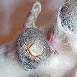
1. A corn on a digital footpad of a retired racing greyhound.
ETIOLOGY
The cause of corns is unknown, but the most popular theory is that the absence of a thick adipose layer in the greyhound footpad allows concussive force between the phalanx and footpad, which damages the skin and footpad.1,2 Another theory is that corns result from footpad cuts or punctures that heal and become fibrous and scarred.1,2 Other theories are that a foreign body or a papillomavirus causes corns to form.1,2
DIAGNOSIS
Corns can usually be diagnosed during a physical examination. In our experience, a dog with true corns typically will be in enough pain that it will be limping. Since the cause of corns is unknown, a radiographic examination of the affected paw should be performed in dogs suspected of having corns to rule out a radiopaque foreign body.
After surgical excision of a corn, a histologic examination should be performed to characterize the microscopic appearance of the lesion, which is usually described as orthokeratotic hyperkeratosis. Use the histologic examination results in combination with antipapillomavirus immunohistochemistry and papillomavirus polymerase chain reaction test results to exclude a papillomavirus as the etiologic agent. In addition, the results of a histologic examination may rule out viral causes of corn formation.

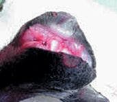
2. A cross-sectional incision on the digital footpad of a retired racing greyhound. Note how deep into the footpad the corn extends.
TREATMENT
The treatment method for corns is as debatable as their etiology. Many techniques have been described. All of these treatment options have varying degrees of success.
Soaking paws and applying manual pressure
We have no experience with soaking the paw and applying manual pressure to express a corn.3 Corns are generally painful with aggressive palpation, and this technique may be more painful and time-consuming than the core removal technique we describe (see "Dental root elevator technique").
Surgical techniques
Various surgical techniques have been described, including corn removal with a laser, scalpel blade, or biopsy punch.3,4 However, a high percentage of corns will return two or three months after surgery.1
Amputating the affected digits is another surgical approach to treating corns. However, even after this radical treatment approach, the risk of corn growth persists in the other digits. It is our opinion that amputation should be performed only when all other treatment modalities have failed.
Silicone injections
Recently, researchers at Auburn University's College of Veterinary Medicine have preliminarily studied whether silicone block gel particles injected subdermally under the distal interphalangeal joint may help provide increased cushioning similar to the fibroadipose tissue of the digit and, thus, prevent the recurrence of corns.1
In this study, some migration of the gel occurred, and some gel remained under the pressure point with evidence of fibrous connective tissue growth around individual gel particles to hold them in place.1 Contact pressure analysis showed a decrease in pressure three months after injection when compared with controls and preinjected pressures.
Flattening, softening, or padding corns
Flattening corns by using a rotary tool to erode or grind them every few weeks reduces load transmission through the footpad and can alleviate some pain. Softening corns can also alleviate pressure and can be accomplished through daily application of a keratolytic agent (KeraSolv gel—DVM Pharmaceuticals) (salicylic acid, sodium lactate, and urea) or over-the-counter corn treatment products for people. In addition, many dogs benefit from avoiding pavement or wearing padded boots (e.g. Thera-Paw—Thera-Paw).
Duct tape
Applying duct tape to lesions has been reported to be effective in treating warts in people.5,6 Corns are grossly similar to warts, but since we do not know what causes corns, we do not know if they are similar in nature to warts in people. Using duct tape to treat canine digital corns has not been studied. In our experience this treatment may benefit some dogs; however, it is difficult to keep the duct tape in place on the corn for a sufficient period of time.


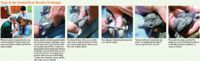
Steps in the Dental Root Elevator Technique
Dental root elevator technique
A corn treatment technique developed by one of the authors (Macherey) is to remove the lesion's hard core with a sharp flat-tipped dental root elevator (see boxed text "Steps in the Dental Root Elevator Technique"). The size of the dental root elevator will vary based on the corn's size. With this technique, dogs usually require no sedation, pain medication, or local anesthesia and may remain standing for the entire procedure. However, dogs that are extremely fearful or aggressive or that are in a lot of pain may require sedation, pain medication, or local anesthesia.
The entire procedure should take less than five minutes. After surgery, monitor dogs for improvement in lameness (most dogs walk out better than they walk in). No physical therapy or medication is required. Over several weeks to months or in as little as three weeks, the corn may return, and the procedure can be repeated.
Carol L. Macherey, DVM
Grassmere Animal Hospital
3926 Nolensville Road
Nashville, TN 37211
William E. Feeman III, DVM
Animal Medical Centre of Medina
1060 South Court St.
Medina, OH 44256
REFERENCES
1. Swaim SF, Amalsadvala T, Marghitu DB, et al. Pressure reduction effects of subdermal silicone block gel particle implantation: a preliminary study. Wounds 2004;16:299-312.
2. Borghese IF. Corns and warts: definitions, causes, and treatments. Celebrating Greyhounds 2003;8:48-51.
3. Andelman NC. Dermatology. In: Bloomberg MS, Dee JF, Taylor RA, eds. Canine sports medicine and surgery. Philadelphia, Pa: WB Saunders Co, 1998;35-44.
4. Blythe LL, Gannon JR, Craig AM. In: Care of the racing greyhound: guide for trainers, breeders and veterinarians. Santa Barbara, CA: Veterinary Practice Publishing Co, 1994;185-229.
5. Lynch TJ. Duct tape removes warts. J Fam Pract 2003;52:111-112.
6. Focht DR III, Spicer C, Fairchok MP. The efficacy of duct tape vs cryotherapy in the treatment of verruca vulgaris (the common wart). Arch Pediatr Adolesc Med 2002;156:971-974.