Veterinary students assist with lion spay in South Africa
See behind the scenes as two South African veterinarians and a group of students and faculty from the University of Missouri's College of Veterinary Medicine attempt to perform an ovariohysterectomy on a lion.
In July 2010, a group of 10 veterinary students and two faculty members from the University of Missouri’s College of Veterinary Medicine traveled to South Africa. Their objective: To experience firsthand the day-to-day lives of two South African veterinarians who work with lions, buffalo, elands, and other animals at private game reserves.
What was the students’ favorite part of their trip? Attempting to spay a lion in the back of a pickup truck.
Click “next” below to read more about this adventure—and learn why the procedure was unsuccessful.


Photo by Rachel Squires
Front row (left to right): Students Lisa Najarian, Kim Doller, Stephanie Carle, and Crystal Garnett
Back row: Student Meagan Brophy, Dr. Ron Cott (associate dean of student and alumni affairs and director of development), Vicki Miller (fiscal manager), and students Anna Stewart, Addie Cable, Rachael Squires, Colleen Risinger, and Josiah P. Sanabria Noriega
< Previous: The team | Next: Darting >
Date: July 22, 2010
Location: Kwandwe Private Game Reserve, situated in the Eastern Cape of South Africa. Kwandwe means “place of the blue crane” in Xhosa, one of the official languages of South Africa.
Assignment: Locate, tranquilize, and transport a female lion back to the Kwandwe work area to perform an ovariohysterectomy.
The MU students and faculty, two South African veterinarians, and a capture team start searching for the lion early in the afternoon. Her transmitter isn’t working, so it takes the team until dusk to locate the lion. As nightfall blankets the African wilderness, the temperature plunges toward freezing.


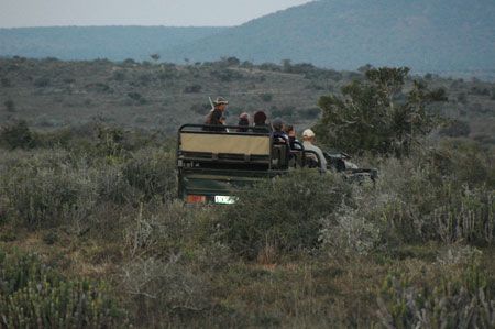
Photo by Dr. Ron Cott
< Previous: The assignment | Next: Vitals >
Using a combination of medetomidine and zolazepam with tiletamine, Dr. Peter Caldwell darts the lion from a Land Rover and watches as she lets out a growl, leaps into the air, and runs out of sight down a hill. The vehicle follows the lion, keeping its distance, until she becomes immobilized.
The Kwandwe capture team loads the 450-lb lion into the back of a pickup truck to transport her to the work area.


Photo by Dr. Ron Cott
< Previous: Darting | Next: Lion's age >
Student Colleen Risinger checks the lion’s vitals on the way into the reserve work area.
After immobilization, the lion is sedated but still sensitive to the external environment, and loud noises or bright lights could awaken her. In addition, the tiletamine causes an initial mydriasis, and any bright light—whether sunlight or artificial light—could cause retinal damage. So lions’ eyes are routinely covered after immobilization.

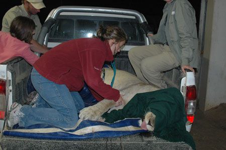
Photo by Dr. Ron Cott
< Previous: Vitals | Next: Students' tasks >
Using teeth to estimate her age, the group established that this lion was 12 to 14 years old. Although the average lifespan varies depending on the environment, lions can live up to 20 years.

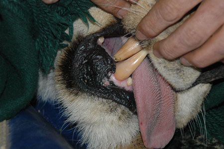
Photo by Dr. Ron Cott
< Previous: Lion's age | Next: Prep >
Each student has his or her own responsibility:
- Administering eye lubricant
- Administering penicillin for the dart wound
- Monitoring intravenous fluids
- Clipping and prepping the surgical site
- Checking the lion’s pulse using a digital artery on top of her paw
- Checking the mucous membrane color, moistness, and capillary refill time
- Checking the palpebral reflex and jaw muscle tone
- Monitoring respirations
- Preparing intravenous drugs for the veterinarians to administer

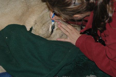
Photo by Dr. Ron Cott
< Previous: Students' tasks | Next: Scrubbing in >
“We clipped, cleaned, and prepped for surgery just like you would for a housecat,” says student Crystal Garnett.
African lions are listed as vulnerable on the International Union for Conservation of Nature and Natural Resources Red List of Threatened Species. So why did the reserve manager request the spay? Lions are allowed to hunt at Kwandwe in an effort to keep them in the most natural wildlife habitat as possible. But to keep the carnivores from overtaking other game, such as antelope, the reserve management had to prevent further population increases. In addition, this female had lived on the reserve long enough that most of the males were her sons, and her advancing age was making pregnancy considerably difficult on her overall health.


Photo by Dr. Ron Cott
< Previous: Prep | Next: Surgery begins >
Peter Caldwell, BSc, BVSc (right), who led the surgery, and Brendan Tindall, BSc, BVSc, scrub in. Dr. Tindall places an intravenous catheter for the delivery of fluids and maintenance anesthesia throughout the procedure.
Although Dr. Caldwell had proposed that the surgery be done off-site in his sterile surgery suite, the reserve management said the cost was prohibitively expensive. Despite the conditions, the veterinarians take every available precaution to keep the workspace sterile.

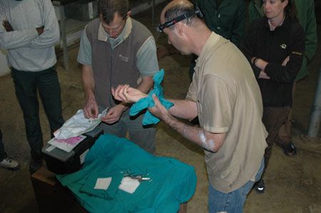
Photo by Dr. Ron Cott
< Previous: Scrubbing in | Next: Restraint >
Dr. Caldwell begins the surgery while students continue to monitor the lion. She is maintained on intravenous propofol and medetomidine and intramuscular injections of zolazepam with tiletamine.
The team soon learns that the lion’s pride had made a kill that day, and her stomach is full of food. Dr. Tindall taps the stomach to relieve the bloating.


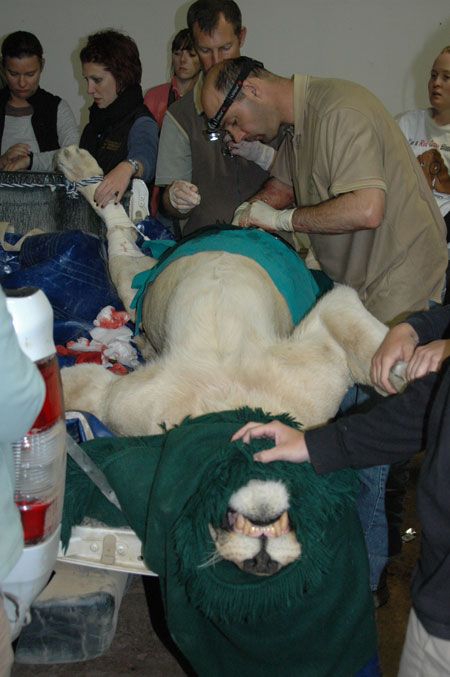
Photo by Dr. Ron Cott
< Previous: Surgery begins | Next: Adhesions >
Two of the lion’s paws are tied down, and students hold the other two.
Twice during the three-hour procedure, the lion becomes light on anesthesia. Student Rachel Squires had just notified the veterinarians that the lion’s heart rate had increased and had already started drawing up propofol to top her off. “I was shaking trying to get the propofol out of the bottle,” she says. “It was intense!”
Dr. Tindall injects the propofol into the catheter and increases the fluid rate. Within seconds, the lion relaxes and the procedure continues.

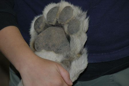
Photo by Dr. Ron Cott
< Previous: Restraint | Next: A second opinion >
As the surgery progresses, Dr. Caldwell discovers multiple adhesions from the lion’s ovaries to her kidneys and body wall. The adhesions prevent him from being able to visualize the uterus and ovaries well enough to remove them.
Dr. Caldwell also discovers three internal transmitters, which likely contributed to the adhesions. He suspects that as the lion moved from reserve to reserve, each location inserted a new transmitter. “As a predator specialist, I would have to criticize a veterinary colleague that does this,” Dr. Caldwell says. He removes the unneeded transmitters.
“I have developed a technique where one can use the omentum to make a pouch and surgically suture the pouch with the inserted transmitter implant in it to the lateral ventral abdominal wall.” Dr. Caldwell adds that this technique:
- Makes the transmitter very easy to find for removal
- Does not require a large incision to remove
- Prevents the transmitter from causing intestinal bruising and damage.

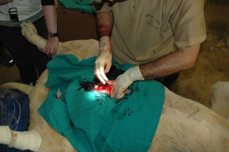
Photo by Dr. Ron Cott
< Previous: Adhesions | Next: Sutures >
Dr. Caldwell requests a second opinion from Dr. Tindall, who agrees that the adhesions are too numerous to exteriorize the uterus and ovaries.
Since the procedure is not successful, the veterinarians decide instead to use a contraceptive on the female. They plan to dart her again in the future and place a microchip-like device by her shoulders that will release a hormone (GnRH) to prevent it from cycling. This method lasts up to 18 months.

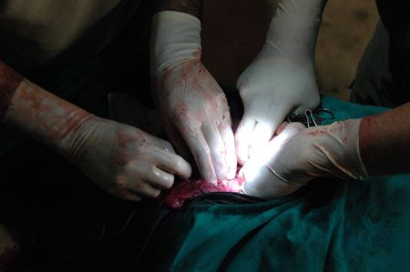
Photo by Dr. Ron Cott
< Previous: A second opinion | Next: Outcome >
Since it is likely the lion or her cubs could pull the sutures out, Drs. Caldwell and Tindall use a three-layer closure with simple interrupted sutures. Because of the stomach bloating, it is very difficult to oppose the edges of the incision.

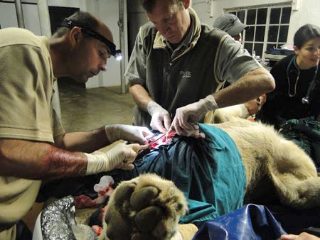
Photo by Stephanie Carle
< Previous: Sutures | Start over >
The lion is loaded back onto the truck, and Dr. Caldwell and a small group travel back out into the field, unload her, and administer the anesthesia reversal agent. Despite looking weak, she is walking and getting around well.
The purple spray visible on the lion’s abdomen is Necrospray (oxytetracycline), which a student had applied both to the incision site and dart wound to prevent infection.
“This was by far the best day of our entire trip,” Garnett says. “It’s not every day you do surgery in the freezing cold on the bed of a pickup in a garage at 9 p.m. at night!” says Garnett.


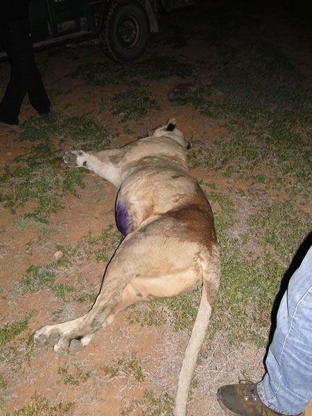
Photo by Stephanie Carle


