Why you need high-quality digital dental radiographs-and how to get them
Looking to make an investment for your practice this year? Put a digital dental radiography unit on your wish list, and you and your patients will all have happy and healthy smiles.


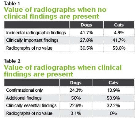
Dental radiography is an essential component in the daily delivery of high-quality dental care for dogs and cats. The diagnostic yield of full-mouth radiographs in feline and canine patients is extremely high, and routine full-mouth radiography is justified. Two studies found that dental radiographs were clinically useful in most canine and feline dental patients (Tables 1 & 2).1,2


Recently, many state-of-the-art veterinary practices have switched over to digital dental radiography because of the speed and ease in which these images can be produced and evaluated. The real value in taking digital dental radiographs is improved patient care while at the same time providing a profit center for the hospital. The advantages of digital dental radiography far outweigh the disadvantages of this new technology (see Advantages and disadvantages of digital dental radiography).
With proper orientation and training in the use of digital dental radiographic units, this new technology can become an integral part of small-animal practices. After an appropriate training period, veterinarians and veterinary technicians will be able to obtain high-quality dental images, which will result in the recognition of more lesions that can then be appropriately treated.
How to take digital dental radiographs
There are two methods of acquiring digital dental radiographs, either digital radiography (DR) or computed radiography (CR). DR images are acquired by placing a sensor into the mouth in the same position as a film and exposing the sensor with a greatly reduced dose of radiation compared with traditional dental film. The image is transferred within seconds for viewing on a computer. These images are then electronically stored and manipulated as needed for radiographic evaluation of a wide variety of dental lesions.
CR images are an indirect way of acquiring digital dental radiographs. With this technology, a reusable phosphor storage plate (PSP) is exposed to X-rays, and the PSP is then processed and converted to a digital image on a computer. CR systems produce a digital image by scanning PSPs of various sizes (0, 2, 3, and 4) that have been exposed to X-rays. These systems allow computer storage, processing, retrieval, and display of the computed radiographic images by using a user-supplied software package. These systems also have an in-line plate eraser function that removes the latest image from the plate immediately after scanning, providing an efficient one-step scanning and erasing process, leaving the PSP ready for collecting the next radiographic image.
Digital dental radiographs can be manipulated for better visualization. The mouse can be used to adjust the contrast and brightness; a particular area of a tooth can be highlighted, magnified, labeled, flipped, rotated, or measured; or explanatory notes can be added.
Indications for taking dental radiographs
Full-mouth radiographs are recommended in every patient, but this may not be possible because of cost constraints or concerns for time under anesthesia in critically ill patients. Digital radiographs can help alleviate these concerns because of the decrease in time needed to acquire digital radiographs.
If full-mouth radiographs are not taken, there are several indications in which teeth should be radiographed. Dental radiography is recommended in the evaluation of:
> Tooth resorption
> Periodontal disease
> Endodontic disease, including discolored or fractured teeth and facial swelling
> Retained roots
> Missing teeth
> Abnormally located teeth
> Malformed teeth
> Osteomyelitis
> Boney lysis secondary to neoplasia
> Metabolic bone disease
> Dentigerous cysts
(localization)
> Traumatic injuries
Dental radiography is indispensable in the development of an appropriate treatment plan.
Positioning for optimal radiographs
Numerous publications describe appropriate positioning for optimal dental radiographs.3-8 There are two specific intraoral radiographic dental techniques: the parallel technique and the bisecting angle technique.
Lessons from CVC
Listen to Dr. Manfra Marretta explain how to easily manipulate digital radiographs for accurate and immediate interpretation.
Parallel technique. The ideal dental radiograph is produced by using the parallel technique. When using this technique, the plane of the radiographic film is parallel to the long axis of the tooth and perpendicular to the plane of the radiographic beam. The parallel technique in dogs and cats can only be achieved with the mandibular premolars and molars. The flat shallow palate and the shallow caudally extending mandibular symphysis in dogs and cats prevent use of this technique when radiographing the maxillary premolars and molars and the incisor and canine teeth.
Bisecting angle technique. For teeth that cannot be captured with the parallel technique, the bisecting angle technique can be used. The film is placed as parallel as possible to the teeth being radiographed. An imaginary line that bisects the angle between the long axis of the tooth and the film is the bisecting angle line. The X-ray beam should be directed perpendicular to the bisecting angle line. Improper utilization of the bisecting angle technique will result in an elongated, a foreshortened, or an overlapped dental image.
How many views? A basic dental radiographic survey consists of six views: the rostral maxillary and mandibular projections, the right and left maxillary projections, and the right and left mandibular projections. Additional radiographs may be necessary depending on the size of the patient and tooth being evaluated. The upper fourth premolar requires additional radiographs to permit adequate visualization of all three roots. A 30-degree rostral oblique projection needs to be added to the bisecting angle technique to permit adequate visualization of the mesiobuccal and palatal roots.
Critiquing dental radiographs
Guidelines have been established to critically evaluate dental radiographs.3 Striving to follow these established guidelines will produce diagnostic films. The following requirements can be used as a guide to assist in self-evaluation of radiographs.
> All teeth to be evaluated are clearly visible.
> The radiographs are
well-positioned.
> The maxillary cheek teeth should have the roots facing upward and the crowns downward.
> The mandibular cheek teeth should have the crowns facing upward and the roots facing downward.
> Maxillary incisors should have the roots facing upward and the crowns downward.
> Mandibular incisors should have the roots facing downward and crowns upward.
> When viewing the right side of the mouth, the anterior teeth are on the right side.
> When viewing the left side of the mouth, the anterior teeth are on the left side of the radiograph.
> Proper angulation has
been used.
> No foreshortening or elongation is present.
> Visualization of all roots and apices is adequate.
> Exposure and developing technique are adequate.
> No artifacts appear on the radiograph.
> Contrast and density of the radiograph are correct.
References
1. Verstraete FJ, Kass PH, Terpak CH. Diagnostic value of full-mouth radiography in dogs. Am J Vet Res 1998:59(6):686-691.
2. Verstraete FJ, Kass PH, Terpak CH. Diagnostic value of full-mouth radiography in cats. Am J Vet Res 1998:59(6):692-695.
3. Holmstrom SE, Frost Fitch P, Eisner ER. Dental radiology. In: Veterinary dental techniques for the small animal practitioner. 3rd ed. Philadelphia: Saunders, 2004;131-174.
4. Bellows J. Dental radiography. In: Small animal dental equipment, materials and techniques: A primer. Oxford, UK: Blackwell Publishing, 2004;63-103.
5. Beckman B. Veterinary dental radiography positioning guide. Available at: veterinarydentistry.net/x-ray-book.
6. Gorrel C. Veterinary dentistry for the general practitioner. 2nd ed. Philadelphia: Saunders, 2014.
7. Lobprise HB. Blackwell's five-minute veterinary consult clinical companion: small animal dentistry. Oxford, UK: Blackwell Publishing, 2007.
8. DuPont GA, DeBowes LJ. Atlas of dental radiography in dogs and cats. St. Louis: Saunders, 2009.