CVC highlight: It's all in the eyes: A look at ocular signs of disease
Dr. Kaese reviews ocular signs that may indicate systemic disease in patients.
Various metabolic, infectious, and immune-mediated diseases as well as hypertension and neoplasia may manifest with ocular signs. Here's a rundown as a reminder when you're looking into your patients' peepers.
Metabolic diseases
Diabetes mellitus. In comparison to dogs, cats have relatively few ocular complications related to diabetes. The most common complication in dogs is the rapid development of bilateral cataracts. Initially, vacuoles form along the equator of the lens, which progress into the anterior and posterior cortex. This rapid cataract development can lead to severe lens-induced uveitis. In addition, diabetic dogs have been shown to be at a higher risk for keratoconjunctivitis sicca and have reduced corneal sensitivity.

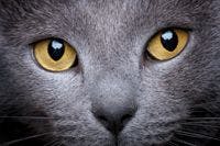

THINKSTOCK/DUSAN MILOVANOVIC
Hyperadrenocorticism. Many ocular lesions have been associated with hyperadrenocorticism in dogs. Surface lesions include progressive and nonhealing corneal ulceration, corneal degeneration, and keratoconjunctivitis sicca. Intraocular lesions include lipemic aqueous humor, lipemia retinalis, and hypertensive chorioretinopathy. Also keep in mind that dogs with hyperadrenocorticism suffer from sudden acquired retinal degeneration more commonly than unaffected dogs do.
Hypothyroidism. Ocular manifestations of hypothyroidism in dogs can result from hyperlipidemia leading to corneal lipid dystrophy, lipemic aqueous humor, and uveitis or lipemia retinalis with retinal bleeding and retinal detachment. Affected dogs are also more likely to suffer from nonhealing corneal erosions.
Infectious diseases
Tick-borne disease. Infection with Ehrlichia canis, Rickettsia rickettsii, and Borrelia burgdorferi can cause conjunctivitis, subconjunctival hemorrhage, anterior uveitis, posterior uveitis, chorioretinitis, retinal hemorrhage, or optic neuritis. However, ocular signs are only found in 10% to 15% of naturally infected dogs.
Histoplasmosis. Ocular signs of this fungal disease include blepharitis, uveitis, chorioretinitis, granulomatous retinal detachment, and optic neuritis.
Toxoplasmosis. Serologic testing indicates that up to 79% of cats with uveitis are seropositive for Toxoplasma gondii. However, there is a high Toxoplasma-positive prevalence in healthy, normal cats, and it's uncommon to find the organism in the uvea of a seropositive cat with uveitis.
Immune-mediated diseases
Uveodermatologic syndrome. This canine disease normally produces bilateral anterior and posterior uveitis, often accompanied by serous retinal detachment. This disease only occurs in an eye with pigment, so it can be unilateral in dogs with one blue and one brown eye. Ocular disease will often precede dermatologic signs of poliosis and vitiligo.
Infectious canine hepatitis. This immune-mediated disease caused by canine adenovirus has become rare since vaccination became routine. Infection or vaccination can cause an interstitial keratitis and uveitis. Ocular signs begin about 10 to 14 days after vaccination. Owners may notice blepharospasm, miosis, and photophobia. Within 24 to 48 hours of the onset of uveitis, corneal edema will develop. The corneal edema can be focal or diffuse and has a ground glass appearance.
Hypertension
Ocular lesions associated with hypertension include optic nerve and retinal edema, retinal vessel tortuosity, retinal hemorrhage, vitreal hemorrhage, hyphema, and retinal detachment. Retinal degeneration occurs secondary to ischemia and retinal inflammation.
Neoplasia
Signs of metastatic neoplasia include conjunctivitis, uveitis, hyphema, and retinal edema. The most common secondary ocular tumor in all domestic animals is lymphosarcoma. In fact, 25% of uveitis is due to neoplasia, and 78% of uveitis due to cancer is lymphosarcoma. Hemangiosarcoma, adenocarcinoma, and transmissible venereal tumors have also been reported to metastasize bilaterally to the anterior or posterior uvea.
Heather Kaese, DVM, MS, DACVIM, DACVO, Eye Care for Animals, Overland Park, Kansas.
Listen in
To hear Dr. Kaese discuss cataract development and the associated complications, as well as treatment options, scan the QR code above, or go to dvm360.com/CVC13Kaese.


")

")


