Skills Laboratory: Cryosurgery for eyelid masses
How to use extreme cold to easily treat tumors on dogs' eyelids.
Next >
Cryosurgery-the removal of tissues by freezing them-is a useful technique for treating eyelid diseases such as benign and malignant tumors and for removing distichiae and ectopic cilia. This article describes how cryosurgery can be used to remove eyelid tumors in dogs.
How cryogens work
Normal eyelid tissue is refractory to damage from freezing primarily because of the tissue's abundant blood supply, which allows for rapid rewarming and necessary nutrition to the tissues. Cryogen-treated tissue is damaged when ice crystals form inside cells. The amount of damage depends on the temperature reached and the rate of thawing of the cells.1,2 In addition, the act of freezing causes secondary inflammation, which augments a lesion's destruction. For most eyelid lesions, two freeze-thaw cycles are recommended. Each cycle should consist of a rapid freeze followed by a slow thaw, as this process produces substantial damage to the lesion.
Several cryogens are used in medicine (e.g. liquid nitrogen, nitrous oxide, and carbon dioxide). Liquid nitrogen is the most popular, effective, and versatile of these cryogens. The boiling point of liquid nitrogen is -196 C (-320.8 F). It is thus able to rapidly freeze tissues to a very low temperature.
Liquid nitrogen can be delivered to the eyelid with a cotton-tipped applicator, a solid cryoprobe, or a spray tip. The cryoprobe is the safest and most useful of these modalities for eyelid surgery as it allows the user to be precise and to avoid spraying liquid nitrogen onto the globe. In addition, cryoprobes come in different sizes (from 1 mm to 6 mm) and shapes (round or rectangular), which adds to their versatility (
Figure 1
).

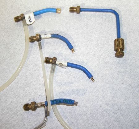
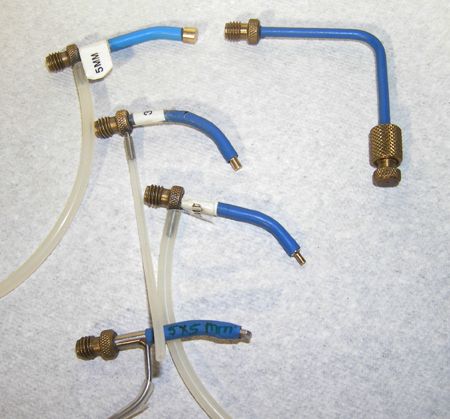
1. Cryoprobes come in many sizes. Shown here on the left are an assortment of 1-mm to 5-mm solid probes and a rectangular probe. On the right is a spray nozzle that fits on the end of the cryosurgical unit and allows for a direct spray of liquid nitrogen to a surgical site.
Cryoprobes attach to handheld liquid nitrogen spray units (Figure 2), which are available from several commercial sources.
Care must be used when handling and applying liquid nitrogen because of its low temperature. Severe overfreezing could slough the eyelid.

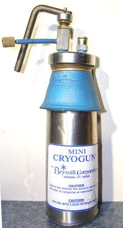

2. Cryosurgical units come in several sizes. The mini-gun, which is light and portable, is good for ophthalmic procedures.
Benefits of cryosurgery
The main advantages of cryosurgery over conventional incisional removal of lesions are as follows:
- In many cases, general anesthesia is not required. The procedure can be performed either with the animal awake or sedated.
- The surgery is relatively noninvasive.
- Large tumors that would require removal of more than one-third of the eyelid can be treated without the need for reconstructive surgery.
- The procedure can be safely repeated.
The most common use of cryosurgery in veterinary patients is to treat benign masses on the eyelids. From 28.7%1 to 60%2,3 of these masses are meibomian adenomas, which are common in aging dogs (Figures 3 & 4).

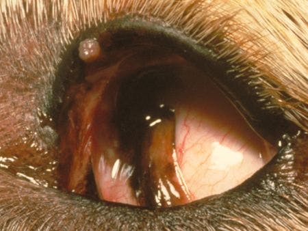
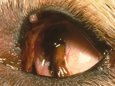
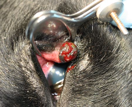
3. The cauliflower-like growth on the upper eyelid of this dog is typical of a meibomian adenoma. These masses are amenable to cryosurgery after excising the bulk of the mass that extends beyond the eyelid margin.


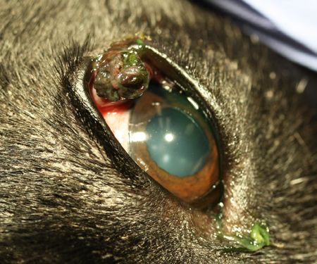
4. A large eyelid growth at the lateral canthus. Even this large tumor is amenable to cryosurgery.
Go to the next page to see the cryosurgery procedure I use to remove masses on the eyelids.
Step 1
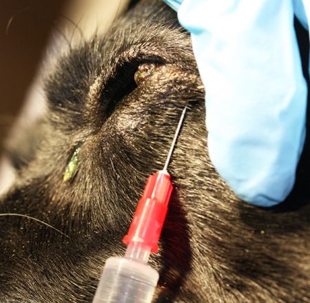
First, manually restrain the dog, usually in lateral recumbency. Thoroughly clean the eyelid three times with dilute (1:4) baby shampoo alternated with rinses with a dilute povidone-iodine solution (not scrub)(1:50 dilution). Then place a few drops of proparacaine (2.5%) on the cornea and palpebral conjunctiva. After applying the topical anesthetic, carefully inject 2% lidocaine into the eyelid above and around the tumor (see photo below). For this injection, I use a 25-ga needle and inject about 0.5 ml in large dogs and 0.25 ml in small dogs.


Step 1 cont'd
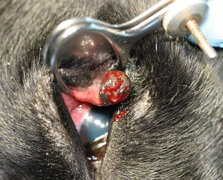
Next, stabilize the eyelid with a chalazion clamp (see photo below). Besides stabilizing the eyelid, this clamp helps control hemorrhage, protects the globe from inadvertent cryonecrosis, and prevents rapid thawing of the lesion. Because the eyelids have an excellent blood supply, clamping them tightly for short periods causes no problems.

Step 2
Using small, blunt-tipped scissors or a surgical blade, excise the tumor flush with the eyelid margin, taking care to maintain the normal architecture of the palpebral margin. As with all masses, place the growth in formalin, and submit it for histologic examination.
After excising the mass, use a small curette to remove any inspissated sebaceous debris. Such debris and possibly inflammation are common because these tumors block the meibomian gland ducts, thus prohibiting the normal extrusion of secretions. If hemorrhage is extensive, place a cotton swab with a drop of phenylephrine on the wound.
Step 3
Before you apply the cryoprobe, a piece torn from the edge of a plastic foam cup may be lubricated and placed under the eyelid to protect the cornea. However, if the chalazion clamp has a back stabilization portion, no other protection is needed.
Freeze the base of the tumor by using a cryoprobe with a diameter about the same as or slightly larger than that of the wound left by the mass excision (see photo below). Place the cryoprobe directly onto the cut surface of the eyelid at the base of the mass, and freeze the area until an ice ball can be seen extending 1 mm from the probe, as seen in the photo. The probe will usually stay frozen to the eyelid after the active freezing procedure has stopped and will come off when the tissue thaws.


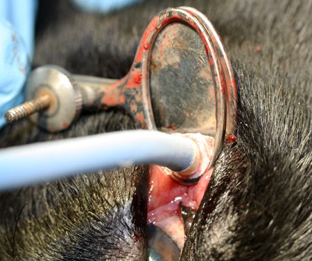
It is important to allow thawing to occur gradually. If the animal moves while the probe is adhered to the eyelid, sterile eyewash can be used to greatly speed up the thawing process and allow the probe to be removed from the eyelid. After the probe is free, allow the eyelid to warm for about one minute and repeat the procedure one time.
< Back
Complications
Complications of eyelid cryosurgery are few. As in every procedure involving tissue excision or incision, infection can occur, but it is rare because of the excellent blood supply to the eyelid. The most common complications are self-trauma, tumor regrowth, and depigmentation of the frozen area. The former can be avoided by placing an Elizabethan collar on the animal and keeping the collar in place for two weeks. Depigmentation is usually temporary, and the tissue usually pigments within six months. However, remember to warn owners about the potential for depigmentation. The prognosis in patients with benign tumors is excellent.
References
1. Andrews MD. Cryosurgery for common skin conditions. Am Fam Physician 2004;69:2365-2372.
2. Krehbiel JD, Langham RF. Eyelid neoplasms of dogs. Am J Vet Res 1975;36:115-119.
3. Roberts SM, Severin GA, Lavach JD. Prevalence and treatment of palpebral neoplasms in the dog: 200 cases (1975-1983). J Am Vet Med Assoc 1986;189:1355-1359.


 in general practice")


")

