Mast cell tumors: new tools in the arsenal (Proceedings)
Mast cell tumors (MCT) are among the most common tumors treated in dogs and are periodically seen in cats.
Mast cell tumors (MCT) are among the most common tumors treated in dogs and are periodically seen in cats. Lesions are typically in the dermis and patients are presented for evaluation of a mass. Diagnosis is easily made in most cases by fine needle aspiration and cytology, with heavily granular round cells distinguished on in-house cytology. Treatment by surgical excision is often successful in general practice. Tumors that recur or are high grade present a challenge and may benefit from referral to a specialist. This talk will include a review of previously known factors in MCT diagnosis and treatment with an updated look at the role of staging and treatment options, as well as insights into novel tests and therapeutics.
Staging
Because canine mast cell tumors spread primarily by lymphatics, staging for this disease parallels that for lymphoma (regional lymph node evaluation, liver, spleen and bone marrow aspiration). Over the years, the value of liver and splenic aspirates has been questioned as it may not correlate well with systemic involvement. This is similar to the discordant results of buffy coat smears in years past. While some practitioners still use buffy coat smears as a monitoring tool with the rationale that if it is positive at diagnosis and clear for mast cells during remission, that buffy coat smears may correlate with relapse. However this has not been shown and buffy coat smears still are often influenced by non-neoplastic diseases. Staging beyond regional lymph nodes was of limited use in a series of dogs with MCT of the muzzle.
It has been suggested that slides created by fine needle aspiration of mast cell tumors may require prolonged fixation for identification of granules. A recent unpublished study found no clear correlation between length of time in the fixative for Diff Quick staining and the ability to diagnose a mast cell tumor by cytologic identification of mast cell granules.
The greatest frustration with staging, including regional and systemic involvement as well as margin identification has been the inability to identify which mast cells are neoplastic and which are innocent bystanders. Recent insights on the molecular abnormalities in canine mast cell tumors may offer new ways to assess mast cells found in the tumor bed, the regional lymph node, the liver, spleen and bone marrow of affected dogs. (see molecular advances below)
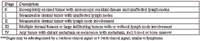
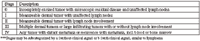
The World Health Organization staging system for canine mast cell tumor is defined below (adapted from Small Animal Clinical Oncology, Withrow and MacEwen, eds., 2001, page 268):

Prognostic factors
The single most important prognostic factor for canine mast cell tumor is the grade of the tumor. Whereas low grade tumors (grade I) have greater than 80% survival in several studies, and approximately 40-50% of grade II tumors survive the first year with surgery alone, grade III tumors treated with surgery alone have poor survival rates of less than 10% long term.
While dogs with multiple tumors (approximately 20-25% of dogs with MCT) are categorized as stage III disease, it has been noted that affected dogs may not have a worse prognosis than lower stages, so long as the disease remains cutaneous. Advanced stage, however is often associated with a worse prognosis and warrants chemotherapy as part of treatment.
Despite earlier studies documenting peripreputial site as a negative prognostic factor, recent studies show that inguinal or perineal location is not necessarily a negative prognostic factor. The tumor grade and behavior (grade 3 and/or recurrent) is more important for these locations. Other mucocutaneous sites including oral and subungual locations as well as visceral MCT continue to be viewed as locations with aggressive tumors.
Another novel concept in the evaluation of MCTs involves the use of proliferation indices. Although grade is considered the most important prognostic factor, markers of cellular proliferation may offer subtle differentiation among similarly graded tumors. These markers include proliferation indices such as AgNOR, PCNA, and Ki-67, and the often mutated receptor tyrosine kinase c-kit. Results have correlated well with grade, subgrade, local recurrence, and metastasis. In multivariate analysis, only AgNOR score and local recurrence were prognostic for survival in a series of nearly 300 dogs. AgNOR staining can be performed on cytology samples suggesting that grade could be obtained on fine needle aspiration alone, but this is tedious and rarely performed. Additionally, attempts to make this correlation have yielded inconsistent results. These tests are commercially available at several laboratories. Of the other proliferation indices, Ki-67 is recognized by the MIB-1 antibody and is expressed in cycling cells. Ki-67 appears to be one of the more promising proliferation markers with more consistent findings among tumors studied. By contrast, PCNA lacks specificity. In one study, Ki-67 helped identify which dogs with grade 2 tumors were more likely to survive. Finally, dogs with tumors that had a mitotic index (number of mitoses per ten high power fields at 400x) of greater than 5 had a median survival time of 3 months, versus 80 months for those with MI less than or equal to 5.
Microvessel density has also correlated well with grade, invasiveness, and survival. Microvessel density is not commonly assessed on routine histopathologic examination and may be influenced by orientation of tumor cells relative to blood vessels.
Some studies report older age as a negative prognostic indicator (>8 years in one study). Additional prognostic factors include growth rate (rapid = bad), and systemic signs (presence = bad). MCTs typically recur or metastasize within the first 6 months after treatment if at all, as indicated by similar short and long-term survival times in some studies.
Molecular advances
Emerging targeted treatments for mast cell tumors are the next great advance in veterinary oncology. Targeted therapies typically exploit some molecular aspect of the cancer cell that is either not found in normal cells or is found with much greater frequency in cancer cells. Stem cell factor receptor, also known as the Kit tyrosine kinase receptor (encoded by the protooncogene c-kit) is important in mast cell tumor growth, proliferation, and differentiation. An activating mutation can lead to constitutive activity in which the cellular signaling is constantly "on" leading to uncontrolled proliferation of mast cells. Activating mutations in the juxtamembrane region encoded by exons 11 and 12 have been found in approximately 30% of canine mast cell tumors and are thought to be spontaneous rather than heritable mutations as they do not appear to occur in the germ line cells. There are, however, breeds such as those of bulldog descent that are overrepresented in studies of canine MCT and these dogs tend to develop tumors that are behaviorally benign. Therefore there may be other genetic influences on the development of this tumor in certain breeds.
New drugs in veterinary medicine have been developed to inhibit c-kit and other tyrosine kinases. Palladia™ (toceranib phosphate) is the first drug that is FDA approved for the treatment of cancer in dogs. The label indication is for recurrent grade 2 or 3 MCT with or without nodal metastasis. The drug was approved in early June 2009 and is currently available to board-certified oncologists and internists until 2010 when full marketing will begin. This was the choice of the company, Pfizer, Inc., to ensure additional field data and experience with the drug before it is released to general practitioners. A series of webinars has been scheduled to educate the veterinary workforce. In addition to inhibition of kit, Palladia™ also inhibits other key split kinases in the same family that are integral to cancer biology, vascular endothelial growth factor receptor (VEGF-R) and platelet-derived growth factor receptor (PDGF-R).
Kinavet® (masitinib) was developed by the European company AB Science, Inc. for the inhibition of c-kit. This company has explored both cancer and non-cancer (atopy) applications for this drug, currently awaiting FDA approval. These drugs are delivered orally and have few side effects, making them an attractive treatment option. In addition, for dogs that have a mutated c-kit gene, the response rate for MCT was very high in some early studies. This is especially important since tumors with kit mutations are more likely to recur and metastasize.
Several laboratories offer tumor evaluation of c-kit status. While identification of c-kit confirms the target of interest prior to treating with an inhibitor, it does not help to predict tumor behavior or patient survival. Likewise, mutation status of c-kit is interesting and may explain successes and failures, but is not necessary prior to treatment, since wild-type c-kit can also be inhibited by these drugs, and dogs with MCT without mutations can also respond accordingly.
Other standard treatment options
Routine cancer therapy consists of surgery, radiation and chemotherapy. Surgery has been and continues to be the initial treatment used. Recent information suggests that low grade tumors may not require the 3 cm margins that have been recommended in the past. If a tumor might be difficult to resect with 3 cm margins, but much more approachable with 2 cm margins, then an incisional biopsy can be helpful to determine the grade prior to definitive surgery. Additionally, a recent abstract reported that only 22% (8/34) of dogs with incompletely excised mast cell tumor had recurrence. Some dogs received prednisone (the effect of which was not quantified), and for those with tumor recurrence, the median time to recurrence was 92 days.
For high grade tumors, chemotherapy is often recommended and vinblastine-containing protocols have performed favorably. Historically vinca alkaloids and alkylating agents in combination with prednisone have been used most frequently. Initial response rates have ranged from 7-40%. By comparison, vinblastine and prednisone therapy yielded a 45% one year survival rate for grade 3 tumors and a 90% survival rate for grade 2 tumors. All dogs that lived to one year went on to live two years. The 90% survival rate with grade 2 tumors is comparable to the control rate with radiation alone, making chemotherapy an attractive alternative to adjuvant radiation therapy. CCNU has also been used for refractory tumors or in conjunction with vinblastine as first-line treatment. In addition to the hepatotoxicity that may occur with CCNU, myelosuppression can be profound and a limited cumulative dose is usually dicatated by waning bone marrow tolerance of the drug.
For improved local control, radiation therapy has traditionally been the treatment of choice. Local control rates exceed 80% in most studies and long-term control is durable. Prior to recommending radiation therapy, it is important to assess the tumor for indications of aggressive behavior (advanced stage or high grade including 'aggressive' grade 2 tumors and grade 3 tumors) as that would warrant chemotherapy. In one study dogs receiving daily fractionation performed better than dogs receiving three times weekly dosing with radiation therapy. In one study of grade III tumors treated with surgery and radiation without chemotherapy, the one year survival rate was 71% and only one of 31 dogs died of systemic disease. Therefore locoregional treatment with radiation may be adequate for some dogs with grade III tumors.
With recent insights on the molecular nature of mast cell tumors, receptor tyrosine kinase inhibitors (RTKIs) may play a role in the treatment of recurrent or refractory MCT. Studies are currently underway to evaluate the efficacy of this class of drugs. In human medicine, gastrointestinal stromal tumors are likewise characterized by c-kit expression, and RTKIs are an example of a therapy targeted to the molecular defect rather than the histologic diagnosis.
Challenges in treating mast cell tumor
When assessing a dog with mast cell tumor, it can be frustrating to determine the best treatment option. Initially local control must be considered. If excision is planned based on cytology alone, then 3 cm margins and one fascial plane deep must be used. If an incisional biopsy reveals a grade 1 or 2 tumors, then 2 cm margins may be used laterally. One frustration is the amount of concern to attribute to the occasional mast cell near the margin or small numbers of mast cells in the draining lymph node. Since mast cells occur naturally in tissues, these may not represent residual disease.
If locally aggressive behavior is demonstrated or predicted, then radiation therapy is an effective adjuvant therapy for MCT. If systemic behavior is a concern, then vinblastine and prednisone chemotherapy with or without an alkylating drug (such as cyclophosphamide or CCNU) should be administered.
In summary, mast cell tumors are a regular part of both general and specialty cancer medicine. Recent advances including molecular characterization and therapy targeting these molecular changes are likely to significantly broaden the treatment options and improve an understanding of prognosis for dogs with mast cell tumors.
References
LaDue T, Price GS, Dodge R, et al. Radiation therapy for incompletely resected canine mast cell tumors. Vet Radiol Ultrasound 1998;39(1):57-62.
Prezoisi R, Sarli G, Paltrinieri M. Prognostic value of intratumoral vessel density in cutaneous mast cell tumours of the dog. J Comp Path 2004;130:143-151.
Thamm DH, Mauldin EA, Vail DM. Prednisone and vinblastine chemotherapy for canine mast cell tumor-41 cases (1992-1997). J Vet Intern Med 1999;13:491-497.
Gieger TL, Theon AP, Werner JA, et al. Biologic behavior and prognostic factors for mast cell tumors of the canine muzzle: 24 cases (1990-2001). J Vet Intern Med 2003;17:687-692.
Hahn KA, King GK, Carreras JK. Efficacy of radiation therapy for incompletely resected grade-III mast cell tumors in dogs: 31 cases (1987-1998). J Am Vet Med Assoc 2004;224:79-82.
Simpson AM, Ludwig LL, Newman SJ, et al. Evaluation of surgical margins required for complete excision of cutaneous mast cell tumors in dogs. J Am Vet Med Assoc 2004;224:236-240.
Sfiligoi G, Rassnick KM, Scarlett JM, et al. Outcome of dogs with mast cell tumors in the inguinal or perineal region versus other cutaneous locations: 124 cases (1990-2001). J Am Vet Med Assoc 2005;226:1368-1374.
London CA, Malpas P, Wood-Follis S, et al. Multicenter, placebo-controlled, double-blind randomized study of oral toceranib phosphate (SU11654) , a receptor tyrosine kinase inhibitor, for the treatment of dogs with recurrent (either local or distant) mast cell tumor following surgical excision. Clin Cancer Res 2009;15(11):3856-3865.