The largest equine corneal transplant ever reported
NC State University ophthalmologists help a show-jumping horse after an eye infection becomes severe

")
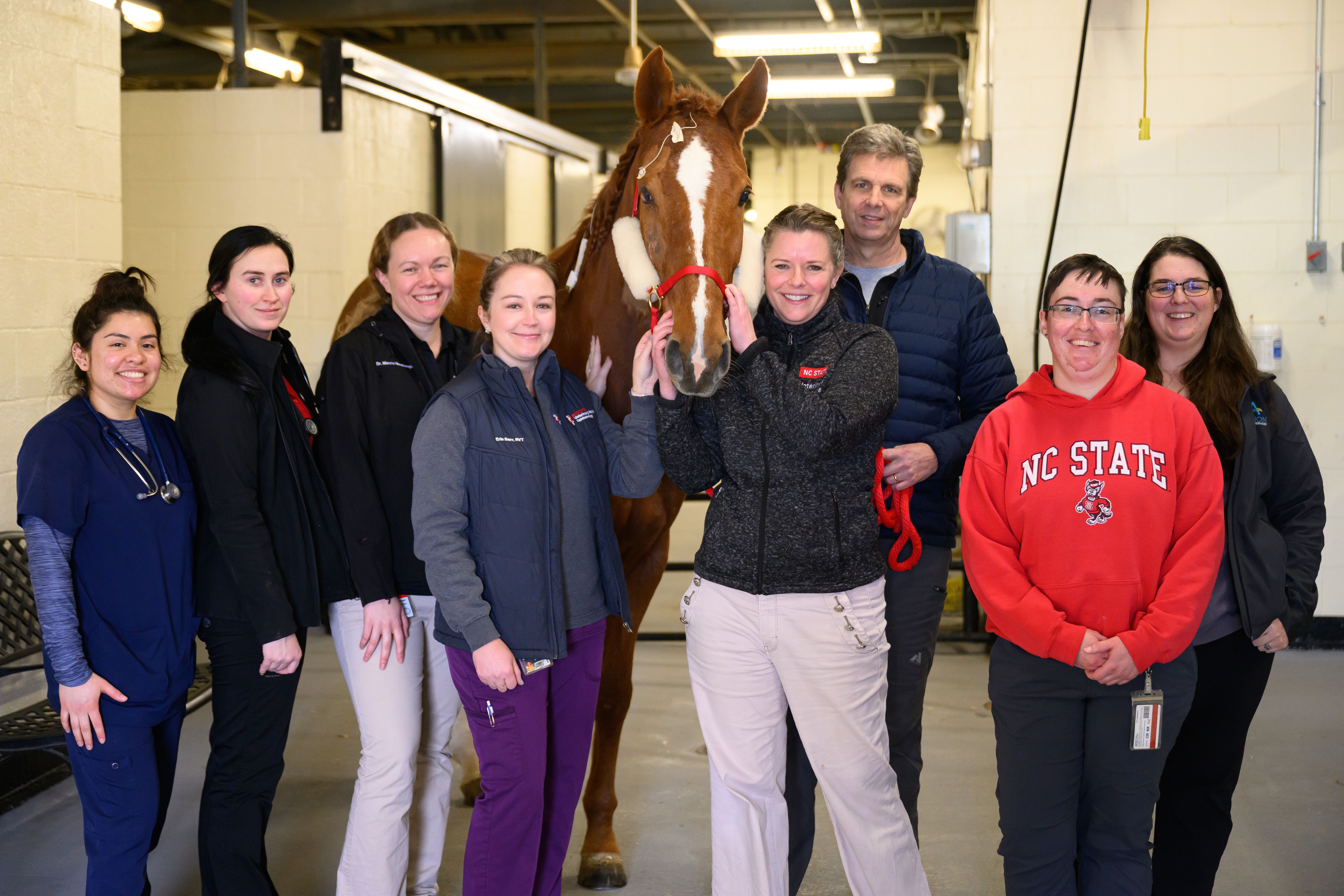
NC State College of Veterinary Medicine ophthalmology and Large Animal Hospital teams with patient, Myra. To Myra's immediate left is Erin Barr, RVT. To Myra's immediate right is Michala Henriksen, DVM, PhD, and Brian C. Gilger, DVM, MS, DACVO, respectively. (Photo credit: John Joyner / NC State College of Veterinary Medicine)
Ophthalmologists from the North Carolina State University College of Veterinary Medicine successfully completed the most extensive resections and corneal transplants ever documented in a horse.1 This groundbreaking procedure involved replacing approximately 60% of the right cornea of a show-jumping Dutch warmblood horse named Myra. Michelle Carnes, MS, DVM, DACVIM (Neurology), veterinary neurosurgeon, and her husband acquired Myra in the Netherlands last fall. Myra developed an eye injury during her relocation from the Netherlands to the United States, unknown to her owners as to how she got the injury. Eventually, the injury led to a corneal infection exacerbated by a fungus that posed a significant threat to her eye.1
As Myra's infection progressed, Carnes sought out help from ophthalmologists across the East Coast and ultimately chose the NC State Veterinary Hospital team. “If anybody was going to be able to save Myra’s eye, it was the NC State team,” Carnes stated in the NC State news release.1
However, before the show horse came to NC State, she was first seen at the University of Georgia’s Veterinary Teaching Hospital, which is closer to her farm in Tryon, North Carolina, and Myra was diagnosed with a treatment-resistant stromal abscess.
“That, as a horse owner, is a devastating diagnosis, because you know that condition can go south really, really fast,” Carnes said.1
Following this diagnosis, Carnes brought Myra to NC State where Brian C. Gilger, DVM, MS, DACVO, professor of ophthalmology and Michala Henriksen, DVM, PhD, associate professor of ophthalmology took on the case at the NC State hospital. The innovative approach NC State took included the application of platelet-rich fibrin (PRF) proteins onto Myra's eye to promote healing.1 The university’s unique research in exploring PRF use for veterinary ocular surgery and the facility’s ultra-high-frequency ultrasound gave Myra the best chance at recovery. NC State is one of few universities nationwide with this advanced ultrasonic device, which lets ophthalmologists see deeper into the eye in more detail than traditional scans allow.
“This ultra-high-frequency ultrasound really transforms how we’re managing these horses’ eyes, because we can see so much better where the disease really is,” Gilger explained. “It allows us to see through tissues that are white or opaque and examine underlying structures, something we cannot do with the usual ophthalmic instruments.”
“Generally, if abscesses are greater than a centimeter in diameter, we know that there’s a very low chance of actually saving the eye, even after surgery. Myra’s measured about 1.4 to 1.6 centimeters,” Gilger continued.1

")
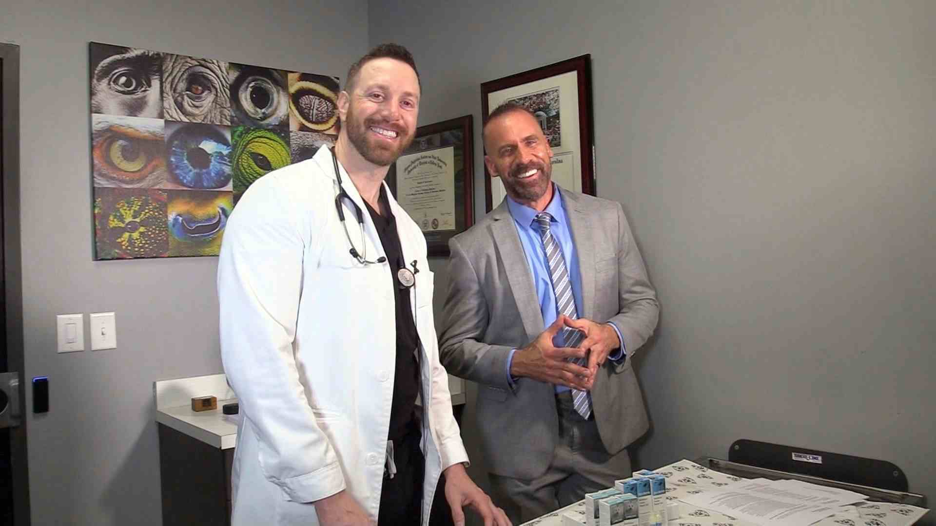
Brian C. Gilger, DVM, MS, DACVO, professor of ophthalmology with Myra. (Photo credit: John Joyner / NC State College of Veterinary Medicine)

To start Myra’s treatment, the ophthalmologist team decided to try antifungal and anti-inflammatory treatments, including luliconazole, a medication that Gilger professionally researches for its use in horses.1,2 This treatment worked at first, and Myra was discharged. Unfortunately, after taking another look at the horse’s imaging, it showed Myra’s abscess resisting treatment and breaking into the anterior chamber of the eye. Myra was quickly scheduled for a surgical resection and corneal transplant.
After removing over half of Myra’s cornea, the veterinary team sutured in cryopreserved cornea from a deceased horse whose owner consented to donation. A conjunctival graft was placed over the corneal graft to protect the horse’s eye from any further infection and to help encourage tissue regeneration and healing.1
With one more challenge thrown their way, Myra’s conjunctival graft unexpectedly detached within 2 weeks, revealing that her cornea had ulcerated underneath. The ulcer showed few signs of regrowing its protective epithelial layer on its own, so the team decided to introduce growth factors to kick-start the tissue repair with the PFR proteins.
Henriksen has studied the clinical use of PRF in dogs with corneal ulcerations and has found it successful in healing the injuries quickly.1,3 “It’s basically tissue glue with healing power, and in Myra’s case, it led to a drastic improvement in her vascularization, or how her blood vessels were growing into the graft, and boosted her scar tissue healing,” Henriksen said.1

")
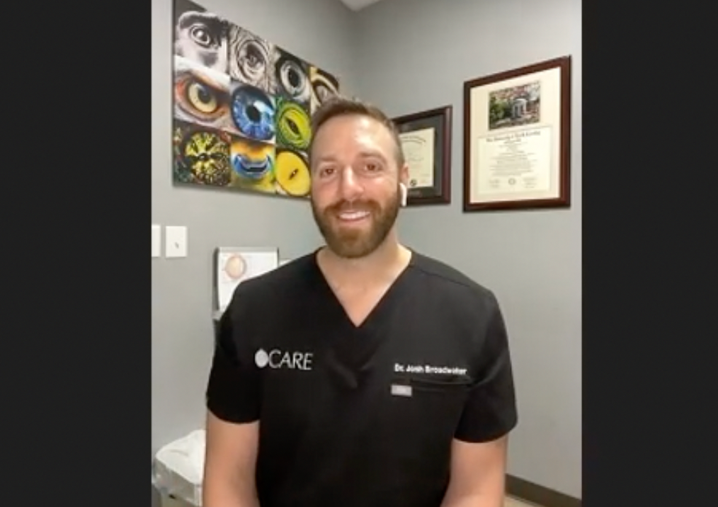
Michala Henriksen, DVM, PhD, associate professor of ophthalmology, with Myra. (Photo credit: John Joyner / NC State College of Veterinary Medicine)

After multiple treatments, Myra is now expected to retain much of her central vision in her right eye but will not recover her hind vision, “like she has built-in blinkers,” Henriksen explained. Although there may be visible opaque scar tissue on her cornea, Myra has received clearance to return home and is fully capable of exercising and competing in jumping events.
“As a person who is in the veterinary profession, I know how difficult it can be sometimes and you get busy, but everybody on the ophthalmology service, the doctors, residents, students, and the technicians, they were all absolutely lovely. And Myra keeping her eye is a testament not only to the expertise of the team, but the unmatched compassion and love they showed her. They continually went above and beyond with Myra’s care and their communication with me. All of their updates made it easier to be away from her. At that point, NC State knew her better than I did,” Carnes said. “She’s doing great on a fitness plan to give her time to build up her strength. Myra loves having so much more space now and has outbursts where she bucks out of joy.”1
“When a horse reaches out to your heart like that, it’s really hard not to get attached. Everybody’s invested, from the treatment technicians to the overnight staff and the people who cleaned her stall. From the ophthalmology team to the whole equine hospital team, it really was just an incredible group effort,” said Erin Barr, RVT, specializing in technician ophthalmology and an NC State hospital employee.1
References
- Rice K. Groundbreaking Transplant at NC State Helps Jumping Horse Stay the Course. News release. North Carolina State University College of Veterinary Medicine. February 20, 2024. Accessed February 21, 2024. https://news.cvm.ncsu.edu/myra-carnes-corneal-transplant/
- Charbonneau M. July 2020 research roundup. North Carolina State University College of Veterinary Medicine. July 23, 2020. Accessed February 21, 2024. https://news.cvm.ncsu.edu/july-2020-research-roundup/
- Baadsgaard Bruun L, Waskiw Hardon TA, Havnsøe Krogh AK, et al. The use of a “horizontal centrifugation protocol” to prepare autologous platelet-rich fibrin membranes for corneal reconstruction surgery in dogs with complicated corneal ulcerations: A case series. Vet Ophthalmol. 2023; 00: 1-15. doi:10.1111/vop.13148


")


