Treatment of forelimb disorders in dogs (Proceedings)
Dogs with strain of the supraspinatus tendon present with a chronic foreleg lameness, some dogs will exhibit periods of non weight bearing lameness.
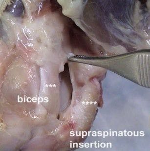
Supraspinatus strain
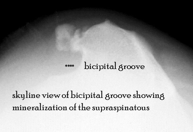
Dogs with strain of the supraspinatus tendon present with a chronic foreleg lameness; some dogs will exhibit periods of non-weight bearing lameness. Although uncommon, any age or breed of dog can be afflicted; however, the condition is seen more in large breeds of dogs (Labrador, Rottweiler). Radiographic views should include standard lateral projections of both shoulders. Mineralization is seen adjacent to the greater tubercle of the humerus. Patterns of mineralization are either irregular, non-homogeneous or well circumscribed and dense foci. A “skyline” view of the bicipital groove is helpful to delineate the location of dystrophic mineralization.


It is worthy to note that the mineralization of the supraspinatus insertion indicating chronic strain may be present but not be the cause of clinical dysfunction. Often mineralization is present in the shoulder in which the dog is not lame. Bilateral mineralization may be present (although different stages of mineralization can be seen between shoulders) and the dog only lame in one shoulder. Recently, MRI has been advocated as a diagnostic tool for shoulder lameness. Some surgeons believe that mineralization of the supraspinatous tendon displaces the biceps tendon causing biceps tendon pain. Although possible, ultrasound examination of the biceps tendon does not demonstrate inflammation secondary to biceps impingement. Also, the position of the biceps tendon is 3-5mm separate from the insertion of the supraspinatous tendon on the greater tubercle of the humerus. Arthrography can be used to outline the bicipital groove to determine if irregularities or filling defects suggestive of bicipital tenosynovitis are present. Diagnosis is based on clinical signs, imaging, and most importantly, ruling out other causes of forelimb lameness. Treatment is enbloc resection of the chronically inflamed section of the tendon. The tendon can be tenodesed in a position where it is exposed to less strain.
Bicipital tenosynovitis

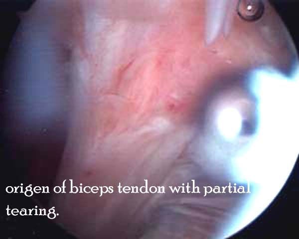


Bicipital tenosynovitis is an inflammation of the biceps brachii tendon and its surrounding synovial sheath. The etiology of bicipital tenosynovitis is either direct or indirect trauma to the bicipital tendon or tendon sheath. Direct trauma due to repetitive injury may be an inciting factor and result in partial or complete tearing of the tendon. Indirect trauma secondary to proliferative fibrous connective tissue, osteophytes or adhesions between the tendon and sheath limit motion and cause pain. It has been hypothesized that mineralization of the supraspinatus tendon causes a secondary mechanical bicipital tenosynovitis. Affected dogs are usually medium? to large?sized, and middle?aged or older. Working and active dogs are more commonly affected; there is no predisposition for either sex. Intermittent or progressive forelimb lameness, which worsens after exercise, is common. The owner may relate the lameness to trauma, but usually there is slow onset of clinical signs. Radiographically, bony resorption at the supraglenoid tuberosity is characteristic of chronic strain at the origin of the biceps tendon. Medical or surgical management have been successful in treatment of bicipital tenosynovitis.
Medical treatment consists of injecting methylprednisolone acetate into the tendon sheath and restricting activity for 3 weeks. In one report (Stobie, JAVMA 1995) reported 50% of dogs treated medically had good to excellent outcome. In the same report, all dogs treated surgically had good to excellent outcome. Given these results, initiating treatment with 1 to 2 steroid injections is a reasonable approach. If outcome is not favorable, surgical intervention is advised. The bicipital tendon may show partial tearing of tendon fibers or partial tearing of the origin of the biceps tendon at the supraglenoid tuberosity. If the biceps tendon shows evidence of partial or complete tearing, the tendon is released by transecting the tendon just distal to the torn portion. Likewise, if there is evidence of synovial proliferation, mineralization, and osteophyte formation within the bicipital groove, the tendon is released from its origin. Tendon release may be performed with either a blade instrument (banana knife, beaver blade, or 11 blade) or a radiofrequency probe. The blade is slightly faster than the radiofrequency probe; however, use of the probe will prevent problems with hemorrhage from vasculature that is often present in the center of the tendon. Upon completion of tendon release, the tendon origin should be closely inspected for small osteochondral avulsion fragments, which can be removed with a motorized shaver or graspers. Lastly, the joint is thoroughly flushed by increasing the ingress flow and allowing egress through a large instrument cannula. Inspect the joint for remaining pathology and then remove the arthroscope and instrument cannula. Suture the portals with non-reactive, non-absorbable suture.
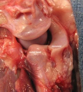
Elbow medial compartment syndrome


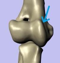
It has been classically attributed to elbow dysplasia. The term canine elbow dysplasia is used to denote an abnormal development of the elbow joint which results in a degree of incongruence. It is used to describe developmental diseases which include ununited anconeal process, fragmentation of the medial coronoid process and osteoarthritis, all believed to be secondary to joint incongruity. The proposed incongruence results in mechanical overload of the medial coronoid/medial humeral condyle through establishment of a “short” radius or “long” ulna. The fact that fragmentation of the medial coronoid will occur with incongruence is well noted in cases of concentric distal radial physeal injury. The latter results in a short radius, mechanical overload of the medial coronoid, and microfracture/fragmentation of the medial coronoid. Does elbow dysplasia (incongruence) exist? Yes. No question elbow dysplasia is seen in younger dogs with developmental incongruence. Not the severe cartilage change and fragmentation of the medial coronoid in the young Irish Wolfhound in the figure.
However, elbow incongruence is not present in all dogs with elbow dysplasia; perhaps we do not have the imaging modality to detect subtle incongruence. Also, elbow dysplasia, a developmental condition that should be present from a juvenile age, does not account for the senior dog who has never experienced a lameness problem and presents with sudden onset lameness. The latter signalment/presentation accounts for the majority of forelimb lameness cases attributed to the elbow encountered by the author. As such, there must be other factors involved in the development of medial compartment syndrome.

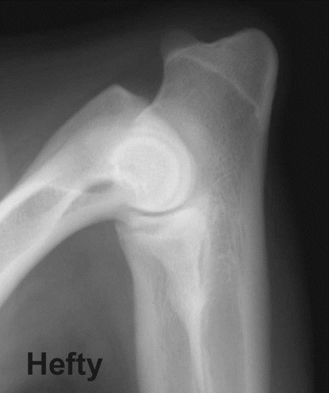


9 yr old Malanois with acute onset lameness: note fragmentation, no articular cartilage pathology
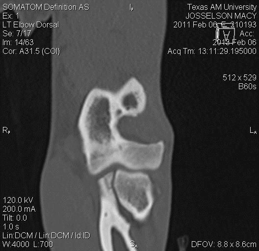
Fitzpatrick (proc ACVS 2010) has shown with the use of micro CT that the pattern of trabecular fracture is consistent with acute traumatic overload (cranial tip fragment) or chronic rotational overload (radial incisures fracture). Recent work by Goodrich (proc VOS 2013) clearly has shown the weight bearing axis of the forelimb to be directed through the medial compartment. Also evident through CT of cases with medial compartment syndrome there increased bone density (sclerosis) in the medial compartment consistent with chronic repetitive mechanical overload. Acute mechanical overload directed through the medial compartment could easily account for the cranial tip fractures as described by Fitzpatrick.


9 year old Retriever with sudden onset lameness; collapse medial compartment
Repetitive mechanical overload generated by years of athletic or playful activity can account for the fragmentation and wear of articular cartilage.


Fitzpatrick has also proposed repetitive rotational overload as a possible cause of radial incisure fragmentation of the medial coronoid. The exact cause of the rotational load is not known but may be attributed to muscular forces (ex. contraction of the biceps/brachialis complex, Hulse Vet Surg 2010) or rotation of the medial humeral condyle against the radial incisure fo the medial coronoid (Bottcher proc ACVS 2013). repetitive rotation of the medial coronoid by contraction of the biceps/brachialis complex compresses the coronoid against the radial head generating a shear stress which corresponds to the fracture plane of a radial incisures fracture


Peter Bottcher has described rotation of the medial humeral condyle
On the radial incisures as a possible cause of fragmentation.
Treatment


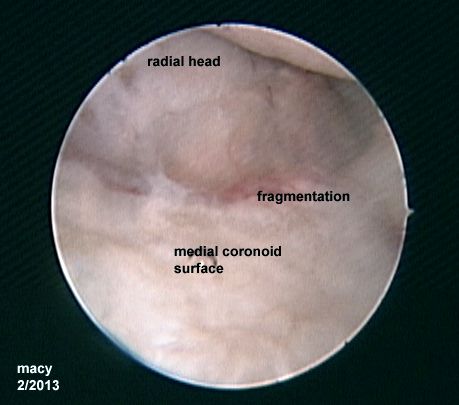
Treatment of medial compartment syndrome includes fragment removal. This includes not only visible fragmentation but fragmentation which may be present beneath the articular surface (subtotal coronoidectomy). If surface articular pathology is present (abrasion) further treatment with medial compartment arthroplasty (abrasion arthroplasty, microfracture), decreasing medial compartment mechanical load (biceps release, sliding humeral osteotomy), and conservative modalities (weight loss, O- 3 FA diet, exercise moderation, nutraceuticals, ACP, stem cell, adequan, polygylcan, NSAIDs) are indicated. If clinical lameness persists, joint replacement (CUE, Tate TER) are considered.
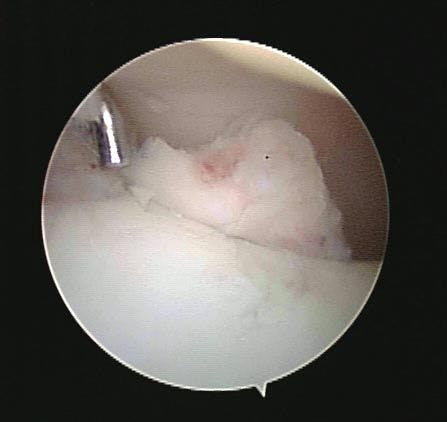
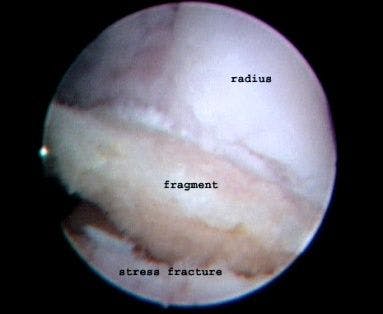
Fragment removal is achieved via arthroscopy or mini-arthrotomy. It is important to note that stress fractures may be present in addition to the obvious visible fragment. A limited subtotal coronoidectomy is advised. A radial incisure fragment in an adult Pittbull with sudden onset lameness. I yr prior to this surgery, a small fragment was removed. Dog never recovered. Repeat arthroscopy found this fragment beneath the articular surface.
Mechanical unloading of the medial compartment:
Biceps ulnar release procedure


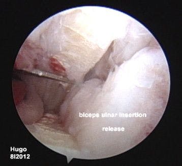

The biceps/brachialis muscles constitute a large muscular complex. The anatomic origin and insertion of the biceps and brachialis muscles are such that the muscular complex exerts considerable force on the medial compartment of the elbow. The force exerted by the biceps is continuous since it is a pennate muscle with central tendon. More importantly, because the insertion of the biceps/brachialis complex is at the ulnar tuberosity, a large polar (rotational) moment is exerted at the cranial segment of the medial coronoid. The magnitude of the polar moment is a product of the moment arm (distance from the ulnar tuberosity to the tip of the coronoid) multiplied by the force created by the biceps/brachialis muscular complex. The polar moment rotates and compresses the craniolateral segment of the medial coronoid against the radial head. The compressive force is medial to lateral transverse to the long axis of the coronoid. A compressive force generates internal shear stress at an oblique angle to the applied compressive force. In this situation, maximal internal shear stress would be oblique to the long axis of the coronoid. Under the right circumstances, the polar moment and resultant compressive force produced by the biceps/brachialis complex may produce sufficient internal shear stress to exceed the material strength of the cancellous bone in the craniolateral segment of the medial coronoid. The result would be microfracture/fragmentation adjacent to the radial head at an oblique angle to the long axis of the medial coronoid. The surgical technique involves releasing the ulnar insertion of the biceps to unload the medial compartment and prevent the rotational moment rotating the coronoid into the radial head.
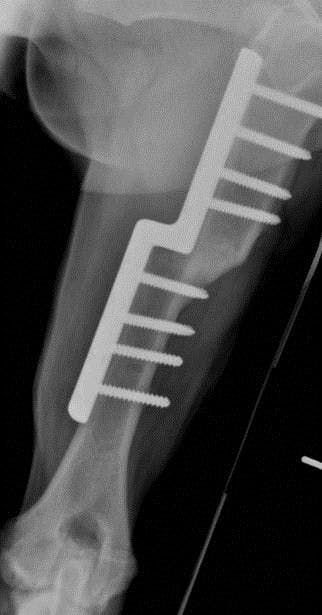
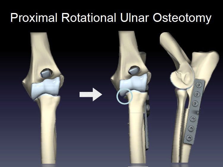
Osteotomies: The deviation of the mechanical axis medially and the humeral varus become more apparent with the progression of medial compartment OA similar to the process in the human knee. The result of overload of the medial compartment, collapse to the medial compartment, and OA. Sliding humeral Osteotomy(SHO): Sliding humeral osteotomy involves creating a midshaft transverse humeral osteotomy and translating (sliding) the diaphysis distal to the osteotomy medially. Doing so shifts the weight bearing axis throught the elbow joint from the medial compartment to the lateral compartment. Owner and vet VAS scores have improved in all cases with a notable decrease in pain upon elbow manipulation


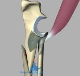
PAUL procedure
The PAUL procedure shifts the weight bearing axis through caudal tipping of the medial coronoid
Dynamic bi-planar ulnar osteotomy described by Noel Fitzpatrick as a method of unloading the medial compartment.
Arthroplasty


Management of articular cartilage lesions is based on the concept that providing blood with mesenchymal cell precursors access to the lesion; this encourages healing by formation of fibrocartilage. Several marrow stimulating techniques have been described to achieve this. Abrasion arthroplasty involves uniform removal of subchondral bone until bleeding is achieved. This can be accomplished in the canine elbow by use of either a curette or burr attachment on a small joint shaver. The shaver is usually more rapid and efficient and generally just as accurate. Another marrow stimulating technique is microfracture. In this technique numerous microcracks are created in the subchondral bone plate with a specialized micropick to allow bleeding at the lesion surface.

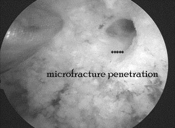


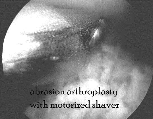

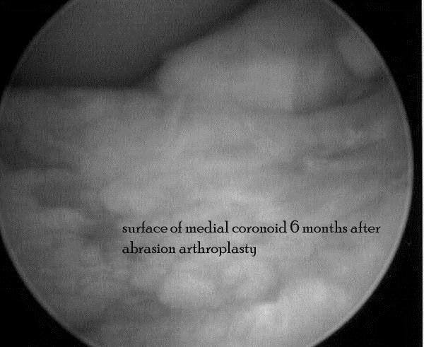
Objective evidence documenting the efficiency of abrasion arthroplasty or microfracture is not available in the dog. The figure to the right shows resurfacing of the medial coronoid in a dog 6 months after abrasion arthoplasty. In man microfracture appears to be more effective than abrasion arthroplasty and is the marrow stimulating technique of choice. The technique is highly dependent on appropriate post operative rehabilitation. In man, 4-6 weeks of non-weight bearing activity coupled with active or passive range of motion is necessary for ideal outcome. Overall, the results of abrasion arthroplasty have been unpredictable and symptoms often recur 2-3 years after surgery. Nevertheless, good to excellent results are reported in 50 – 60 % of patients.


Elbow replacement is an option in dogs which have end stage elbow OA and conservative/less invasive surgical modalities have not resolved clinical pain. A number of prosthesis are available but the most popular one today is the TATE elbow. Clinical outcome studies indicate that a mechanical lameness may pesisit but that the dogs appear to be less painful. A prosthesis presently in clinical trial is the CUE (canine unicompartmental elbow). The concept is simple and carries little morbidity. Information concerning this technique will be forthcoming in the near future.

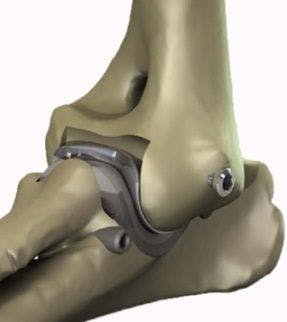

TATE total elbow

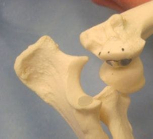

CUE


 in general practice")


