Dental Corner: Diagnosing and treating chronic ulcerative paradental stomatitis
Chronic ulcerative paradental stomatitis (CUPS) is a painful condition in dogs that is also known as ulcerative stomatitis, idiopathic stomatitis, and lymphocytic-plasmacytic stomatitis.
Chronic ulcerative paradental stomatitis (CUPS) is a painful condition in dogs that is also known as ulcerative stomatitis, idiopathic stomatitis, and lymphocytic-plasmacytic stomatitis. The hallmark lesion is a paradental, or so-called kissing, ulcer.1 Patients with CUPS are usually inappetent or anorectic, and the chief complaint from owners is usually fetid halitosis and drooling. CUPS can affect any dog, but Maltese are overrepresented, and a familial predilection has been shown (Figure 1). Cavalier King Charles spaniels also seem to be genetically predisposed.2 While dogs of any age can be affected, I have rarely seen CUPS in patients under 1 year old.


Figure 1 : The classic presentation of CUPS: A Maltese that refuses to eat and whose breath is affecting every room in a three-story house.
Pathophysiology
CUPS lesions are characterized histopathologically by a predominance of lymphocytes and plasmacytes, indicating that the disease is inflammatory rather than infectious. The presumed antigen stimulating the inflammation is bacterial plaque,3 or in terms that pet owners can understand, the dogs become allergic to the bacteria that live on their teeth. Some veterinary dentists refer to this condition as plaque intolerance or plaquehypersensitivity.
As the inflammatory condition continues, gingivitis and advanced periodontitis (deep pocket formation, gingival recession, periodontal bone loss, furcation exposure) can occur concurrently. The change from predominantly gram-positive to predominantly gram-negative bacterial flora that occurs with periodontitis seems to fuel the inflammation of CUPS, suggesting to me that antigens from the gram-negative bacteria may trigger an even stronger inflammatory response in affected dogs.
The severe pain dogs experience from CUPS lesions makes it difficult for owners to provide adequate dental home care. This lack of home care and the patient's unwillingness to chew allow more plaque to accumulate, and the vicious cycle continues.
Clinical signs
Affected dogs exhibit various clinical signs including halitosis (sometimes severe), inappetence or anorexia, ptyalism, and oral pain (pawing at mouth, chattering jaw movements). Other signs may include abnormal chewing movements, eating difficulties, difficult prehension, and oral hemorrhage. Affected dogs often will not chew on their toys and will refuse the hard portions of their diets.

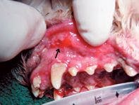

Figure 2 : A paradental, or kissing, ulcer (black arrow) on the oral mucosa apical to the right maxillary canine tooth. Note how this tissue will contact the tooth surface when the mouth is closed.
On oral examination, gingivitis, stomatitis, and, sometimes, advanced periodontitis are observed. The classic distribution of the stomatitis is paradental (Figure 2). The lesions on the oral mucosa correspond with areas that touch the teeth's surfaces. The lesions are red, sometimes slightly raised, and often ulcerated. In addition to the buccal mucosa, the lateral mucosa of the tongue is often inflamed and ulcerated where it touches the lingual mandibular teeth's surfaces (Figure 3). The most severe lesions are often found associated with areas of gingival recession. Concurrent sublingual granuloma, or gum chewers lesions, may exacerbate the problem on the lingual mandibular mucosa. Sometimes, dogs with CUPS will have a concurrent lip fold dermatitis (intertrigo) adding to the oral discomfort as well as an overall foul odor. The connection between these two conditions is most likely that excessive drooling from the CUPS contributes or exacerbates the lip fold dermatitis. Another finding consistent with CUPS is mandibular lymphadenopathy.


Figure 3 : Gingivitis, stomatitis, cheilitis, and inflammation along the tongue's lateral border. To diagnose CUPS, oral biopsy samples of affected areas need to be evaluated.
Initial examination and diagnostic tests
When patients are presented for evaluation of clinical signs of CUPS, perform a complete physical examination. Carefully examine the mandibular lip folds for evidence of intertrigo. Next, perform a complete blood count (CBC), serum chemistry profile, and urinalysis. Other preanesthetic diagnostic tests, such as thoracic radiography and electrocardiography, may be included as well. The only laboratory findings commonly associated with CUPS include hyperproteinemia secondary to a hypergammaglobulinemia (polyclonal) and mild (up to 25,000/ml) neutrophilic leukocytosis. If other abnormalities are encountered in the diagnostic workup, address them as additional problems.
Diagnosis
The differential diagnoses in patients with signs characteristic of CUPS are listed in Table 1. Although CUPS can be presumptively diagnosed based on a patient's history and oral examination findings, a definitive diagnosis can only be made by histologically examining lesions. Anesthetize the patient, and use a 0.3- to 0.6-mm skin biopsy punch to obtain samples; try to obtain at least two samples from the representative areas of the oral cavity. Additionally, perform a thorough oral examination, including charting pocket depth, and note the results on the patient's dental chart. Dental radiography is important in identifying advanced periodontal disease, and a full-mouth series is warranted.

Table 1 : Differential Diagnoses in Dogs with Signs Typical of CUPS*
Once you've definitively diagnosed CUPS, discuss the long-term prognosis with the owners. This condition can usually be treated but will require lifelong therapy or total mouth extractions.
Treatment
The initial treatment plan should include a complete dental prophylaxis: gross calculus removal and ultrasonic crown scaling, subgingival plaque and calculus removal, polishing, gingival sulcus irrigation with chlorhexidine solution, and a thorough oral examination including periodontal probing. It is difficult enough to save the healthy teeth in dogs with CUPS, but it is nearly impossible to treat the diseased teeth and keep the inflammatory disease in check. With this in mind, it is recommended to extract any teeth with advanced-stage periodontal disease at the time of the initial prophylaxis and oral biopsy. Patients with CUPS (or presumptive CUPS) are excellent candidates for OraVet (Merial), which is a waxy barrier applied to tooth surfaces after prophylaxis to retard plaque accumulation.
The two main goals when treating CUPS are to provide immaculate oral hygiene and to achieve clinical resolution by using anti-inflammatory or immunosuppressive medications. The better the level of home care, the fewer drugs needed. Brushing a patient's teeth and applying chlorhexidine daily, applying OraVet weekly, and instituting long-term intermittent antibiotic therapy (pulse therapy) are all home-care tools that can be combined to obtain maximal oral hygiene.
The choice of antibiotics to treat oral infections is based on our knowledge of the common pathogens. Do not perform oral bacterial cultures because more than 200 species of microorganisms inhabit the oral cavity, and the results will only confuse the issue. Antibiotic selection should be based on each agent's ability to affect gram-negative anaerobic bacteria. The short list includes amoxicillin trihydrate-clavulanate potassium, clindamycin, metronidazole, and the tetracyclines. These antibiotics differ in respect to their being bactericidal vs. bacteriostatic, their tendency to cause gastrointestinal side effects, and, in the case of the tetracyclines, their tendency to stain the teeth of actively growing dogs. Combination therapy, such as metronidazole and clindamycin, may be most effective in severe cases. Pulse therapy remains a controversial issue with no good scientific studies to document its effectiveness. However, there are many anecdotal reports of success. The risks of creating resistant bacterial organisms must be weighed against the benefits to a patient. If you choose pulse therapy, a common dosing regimen is 2.5 mg/lb (1.13 mg/kg) clindamycin given twice a day for five days every month.
Prednisone is the drug of choice for suppressing the inflammation associated with CUPS. Start at an anti-inflammatory dosage of 1 mg/kg given twice a day, and taper the dose by cutting it in half every five days until the lowest every-other-day dose is achieved. When the prednisone dose is insufficient, the owners will report a recurrence of clinical signs, and the dose will need to be increased.
If patients do not tolerate prednisone, azathioprine can be used to control the inflammation. Start azathioprine at a dosage of 1 to 2 mg/kg given once a day, and if effective, taper the dosage to 1 mg/kg given every other day after three weeks. Additional monitoring for patients receiving azathioprine includes frequent CBCs. If evidence of bone marrow depression is observed, discontinue the azathioprine, and explore other treatment options.
In patients in which medical therapy is not successful or not advisable, healthy teeth can be extracted to achieve beneficial results.1,2 In some cases, extracting the caudal teeth (molars and premolars) will be enough. In patients in which caudal teeth extraction fails to achieve clinical remission, extract the remaining teeth. In my experience, CUPS almost always resolves when all of a dog's teeth have been removed. Dogs with no teeth can eat and live normal lives. But keep in mind that often, after all the teeth are extracted, a dog's tongue will hang out of its mouth.
Conclusion
CUPS is a chronic condition that requires lifelong patient monitoring. Always stress to owners that this condition will most likely require a lifetime of therapy. If owners run out of their dogs' medication, clinical signs of CUPS commonly recur. A cure may be achieved, however, by extracting many or all of the teeth. Because of the potential side effects from the medications used to treat CUPS, periodic physical and oral examinations should be accompanied by appropriate hematologic tests. Routine dental prophylaxis (every four to six months) should be part of a complete oral hygiene program when the goal is to save teeth.
REFERENCES
1. Harvey, C.E.; Emily, P.P.: Oral inflammatory and immune-mediated disease. Small Animal Dentistry. Mosby-Year Book, St. Louis, Mo., 1993; pp 145-155.
2. Wiggs, R.B.; Lobprise, H.B.: Clinical oral pathology. Veterinary Dentistry Principles and Practice. Lippincott Williams & Wilkins, Philadelphia, Pa., 1997; pp 104-139.
3. Smith, M.M.: Oral and salivary gland disorders. Textbook of Veterinary Internal Medicine, 4th Ed. (S.J. Ettinger; E.C. Feldman, eds.). W.B. Saunders, Philadelphia, Pa., 1995; pp 1084-1097.
"Dental Corner" was contributed by Daniel T. Carmichael, DVM, DAVDC, The Center For Specialized Veterinary Care, 609-5 Cantiague Rock Road, Westbury, NY 11590.