Image Quiz: A Chihuahua with traumatic bite wounds
Correct answer: Image Quiz: A Chihuahua with traumatic bite wounds
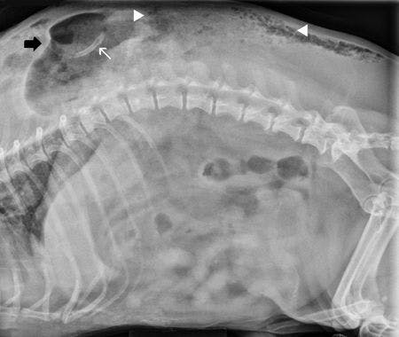
A fragment of the left 13th rib is correct!


Radiograph interpretation
A large gas-filled structure with a distinct wall on the cranial edge (black arrow) is present dorsal to the 10th through 13th thoracic vertebrae (see figure above). The large structure summates with the left caudal thorax on the ventrodorsal view. Subcutaneous emphysema is present (between white arrowheads) dorsal to the caudal thoracic and lumbar vertebral column. The margin of the left caudal thoracic and cranial abdominal body wall is indistinct. There is mildly reduced serosal detail in the abdomen. The stomach and spleen are not identified on the lateral view. The left 13th rib is cranially angulated and fractured. The distal segment is displaced dorsally and is adjacent to the large gas-filled structure (white arrow). No abnormalities of the diaphragm are perceived. The radiographic diagnosis is traumatic body wall hernia with gastric and splenic displacement and fracture and luxation of the left 13th rib.
Case outcome
Exploratory surgery was performed via a ventral midline celiotomy. On initial evaluation of the abdomen, the diaphragm appeared intact. The stomach was herniating through a defect in the left body wall along with most of the spleen.
The remaining abdominal organs appeared normal. The stomach was replaced into the abdomen, and the spleen was excised because of compromised vascular supply. The internal aspect of the body wall defect was repaired by apposition of the muscular layers of the body wall with 3-0 absorbable suture material.
The patient became hypoxemic during the closure of the initial celiotomy. The abdomen was reopened, and a rent in the left side of the diaphragm was discovered, which had led to secondary pneumothorax. Chest tubes were placed, and the diaphragm was repaired using 3-0 absorbable suture material.
The external wounds were lavaged, débrided, and closed routinely. However, the patient was unable to ventilate independently after surgery. Because of the poor prognosis for recovery, the owners elected humane euthanasia. No necropsy was performed per the owners' request.
Discussion
A traumatic body wall hernia is defined as a trauma-induced defect in the body wall, leading to displacement of internal organs outside of the abdomen or thorax.1 Blunt force trauma damages the body wall because of increased internal pressure while the abdominal muscles are contracted, leading to rupture from their attachments.2-4 Shearing forces across a bony prominence may also contribute to tearing of abdominal musculature and protrusion of internal organs.3,5
The skin is more elastic and will stretch across the injury and often will have minimal damage, whereas the deeper, less elastic tissues will be torn.6
One review found the most common locations of traumatic body wall hernias to be the ventral caudal abdomen and the paracostal area,4 while a more recent report found the most common locations to be the lateral paralumbar body wall and rupture of the cranial pubic ligament.6
Bite wounds, as in this case, account for 54% of canine and 40% of feline traumatic body wall hernias, while the remaining are almost always associated with vehicular trauma.6 The most common location of body wall damage due to bite wounds is the dorsal or lateral body wall, but with sharp trauma, herniation may occur anywhere.1,5
A unique aspect of bite wound trauma is the shearing forces when a larger dog picks up a smaller dog and shakes it during the attack. This tears the intercostal muscles and the origins of the external abdominal oblique and transversus abdominis muscles as they stretch,6 which, in this patient, likely contributed to the displacement of the stomach and spleen through the defect in the body wall.
This case also highlights the fact that as many as 75% of animals with abdominal hernias also have concurrent injuries,1,7 as a diaphragmatic rent was discovered on exploratory laparotomy but was not evident on physical examination or imaging. A thorough physical examination and meticulous exploration during laparotomy are strongly recommended for all patients that have sustained traumatic body wall hernias.
Reasons for emergency celiotomy include strangulation or incarceration of abdominal structures, damage from tearing or obstruction of the vascular structures, and sharp trauma resulting in penetration of intra-abdominal or thoracic structures. In general, patients with penetrating abdominal trauma should undergo emergency surgery after stabilization.
References
1. Read R. Traumatic and incisional hernias. In: Slatter DH, ed. Textbook of small animal surgery. Philadelphia, Pa: WB Saunders Co, 1993;443-447.
2. Fossum TW. Umbilical and abdominal hernias. In: Fossum TW, ed. Small animal surgery. St. Louis, Mo: Mosby, 1997;184-187.
3. Damschen DD, Landercasper J, Thomas CH, et al. Acute traumatic abdominal hernia: case reports. J Trauma 1994;36(2):273-276.
4. Waldron DR, Hedlund CS, Pechman R. Abdominal hernias in dogs and cats: a review of 24 cases. J Am Anim Hosp Assoc 1986;22:817-822.
5. Smeak D. Abdominal wall reconstruction and hernias. In: Tobias K, Spencer J, eds. Veterinary surgery: small animal. St. Louis, Mo: Elsevier Saunders, 2012;1353-1380.
6. Shaw SR, Rozanski EA, Rush JE. Traumatic body wall herniation in 36 dogs and cats. J Am Anim Hosp Assoc 2003;39(1):35-46.
7. Dubois PM, Freeman JB. Traumatic abdominal wall hernia. J Trauma 1981;21(1):72-74.